
 metals · microbes · host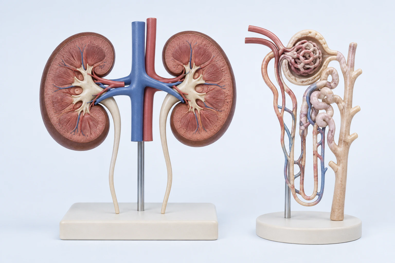
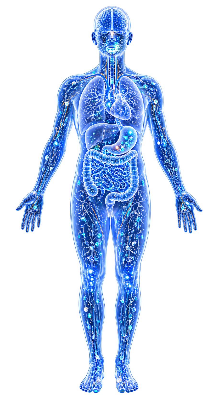
Renal and nephron orientation for chronic kidney disease. CKD spans multiple causes and stages and has no single universal gross appearance; this reconstruction is not a scan, histology slide, laboratory result, severity grade, or diagnosis.
Scientific media record1 verified identifier
- Subject
- Renal Insufficiency, Chroniccondition
- Identifiers
- MeSH:D051436
- Review
- Editorial review completeIdentifiers authority-verified · Accessibility validated · · chronic-kidney-disease|chronic-kidney-disease-pathology-v1.webp
- Digital source
- Trained-algorithmic mediaCreated with a trained generative algorithm and reviewed by WikiBiome for subject identity, scientific framing, identifiers, provenance, and accessibility.
- License
- CC BY-SA 4.0Created
A progressive loss of kidney function affecting approximately 850 million people worldwide (10–14% of adults), causing 1.2 million deaths annually.
CKD is unique among diseases in this wiki because it occupies both sides of the metal-disease equation: Heavy Metals (cadmium (Cd), lead (Pb), mercury (Hg), arsenic (As)) directly cause nephrotoxic injury, AND kidney dysfunction impairs metal excretion, creating a vicious cycle of accumulation and damage.
Beyond metals, two independent lines of Mendelian randomization evidence now establish that specific gut and oral microbiome taxa are causally linked to CKD risk and progression.
Evidence map110 cited passagesInspect provenance +
As CKD progresses and GFR decreases, the ability to eliminate environmental toxicants declines, creating a vicious cycle of metal accumulation and further kidney damage.
CKD patients have +0.23 µg/dL higher blood Pb with simultaneously lower urinary Pb excretion (-0.16 ng/mL), confirming reduced elimination capacity.
Each 10 mL/min per 1.73m² lower eGFR is associated with 0.05 µg/dL higher blood Pb and 0.02 µg/L higher blood Cd.
Reverse causality was directly demonstrated: mixed metal exposure showed a negative joint effect on CKD risk in BKMR modeling, likely reflecting impaired renal excretion.
Cd increases CKD risk from 10% to 25% in exposed individuals.
Blood Cd 1 mcg/L associated with CKD and albuminuria.
Cd impairs electron transport chain complexes II/III, induces ER stress, disrupts autophagy.
Cd specifically targets the proximal tubule, where it accumulates bound to metallothionein.
In the gut microbiome, Cd exposure at low doses specifically decreases Akkermansia muciniphila, a gut barrier-protective species, and selects for cadmium-resistant Proteobacteria carrying cadA resistance genes.
Higher blood Pb is consistently associated with lower eGFR.
Racial disparities: Black race dramatically modifies the Pb-CKD association—10 mL/min per 1.73m² lower eGFR associated with 0.13 µg/dL more Pb among Black participants vs 0.03 among White (4x stronger association).
Black individuals have higher rates of iron deficiency, CKD, and lead susceptibility, compounding vulnerability.
Low-level Pb and Cd exposure associated with increased CKD mortality.
Hg disrupts mitochondrial membrane potential, triggers oxidative stress, causes cytoskeletal alterations, inhibits Na+/H+ exchangers and aquaporin-1.
Kidneys with reduced renal mass are more susceptible to Hg toxicity.
As increases ROS production, activates MAPK/NF-kB pathways, enhances myeloperoxidase activity, induces apoptosis.
Blood arsenic was significantly higher in CKDu patients (91.97 mcg/L) compared to CKD (4.5 mcg/L) and healthy subjects (39.01 mcg/L).
As independently associated with CKDu on multinomial regression (OR 1.013, p=0.014).
In Taiwan's Changhua County (electroplating-contaminated), soil-based Factor 1 metals chromium, copper, nickel, zinc associated with increased ESRD risk (aHR 1.08, p = 0.02).
Only Zn (aHR 1.08) and Ni (aHR 1.08) were individually significant risk factors for ESRD progression.
CKD patients progressing to ESRD had higher residential soil Cu, Ni, and Zn concentrations.
alpha klotho mediates the Hg-CKD association with 34.55% mediation proportion.
Mendelian randomization confirmed higher alpha-klotho levels causally associated with reduced CKD risk (OR 0.9842).
Klotho functions discussed by the source include antioxidant enzyme regulation (SOD, CAT, GPX-4), TLR4 signaling suppression, NF-kappaB inhibition, autophagy promotion, and Ca/vitamin D homeostasis.
Showing 24 of 110 evidence-bearing passages. Every remaining citation is still indexed in the reference record below.
One disease. Five evidence layers.
A generated systems view of the metals, organisms, host sequestration signals, ecological conditions, and microbial functions indexed for Chronic Kidney Disease.
Evidence layer
Taxonomic signature
Organisms reported as enriched or depleted, with their indexed functional context kept beside the name.Urease producer, LPS source, indole producer, siderophore iron scavenging — causal MR evidence for CRF risk (OR=1.22)
Aromatic compound metabolism; uremic toxin (IS, PCS) production; enriched with CKD progression
Proteolytic, urease-positive; indole/p-cresol producers; bloom in uremic environment
Causally increases CKD risk (MR OR=1.15, Bonferroni-significant); H2S-producing sulfate-reducing anaerobes driving systemic inflammation and cholesterol absorption
F. plautii enriched with CKD progression; flavonoid degrader
Multiple species positively associated with CKD severity
Indole and p-cresol producers; proteolytic fermentation
Methanogenic archaea; absent in early CKD, present in ESRD
Enriched in CKD stages 3-5D; proteolytic fermentation producing uremic toxins (indoxyl sulfate, p-cresyl sulfate)
Pathogenic members enriched across CKD stages; uremic toxin producers
Enriched in CKD; includes proteolytic members generating uremic toxins
Enriched in CKD stages 3-5D; paradoxically elevated despite conventional beneficial status
Metal-resistant pathobiont; dominant in CKD stage 3 fecal cultures under metal-selective conditions; produces p-cresyl sulfate
Major butyrate producer; anti-inflammatory; depleted across 4+ studies
Depleted from CKD stage 3b; butyrate producer; loss drives barrier dysfunction
Earliest Lachnospiraceae signal — depleted from CKD stage 3a; butyrate producer
Depleted from CKD stages 5-5D; butyrate producer; persists depleted through hemodialysis
Plant polysaccharide degrader; depleted across multiple studies
Mucin producer; gut barrier protection; specifically depleted by cadmium exposure
SCFA producer; heavy metal binder; negatively correlated with progression (variable in some studies)
Key butyrate species; depleted in CKD
Resistant starch degrader; main contributor to integrated disease network; keystone species
Depleted from CKD stage 3b; butyrate/acetate producer
Depleted from CKD stages 4-5; butyrate producer; enriched by probiotic copper (Cu)/nickel (Ni) detoxification
Five genera consistently depleted (MR confirms causal protection: UCG-010 OR=0.89); the Lachnospiraceae collapse is the defining microbiome event in CKD
Evidence layer
Nutritional immunity
Host metal-withholding, inflammatory, antioxidant, and microbial-metabolite signals indexed in the signature.Elevated host signals
18Depleted protective signals
12Evidence layer
Ecological state
The environmental conditions that connect the organism-level observations into a system.Evidence layer
Virulence functions
Microbial structures, enzymes, and acquisition systems implicated by the linked evidence.The disease record, in full.
The original WikiBiome disease narrative remains intact beneath the generated signature atlas.
Metallomic Signature#
The Metal-Disease Matrix: A Cross-Source Synthesis identifies CKD's profile as: iron (Fe) ↑↓ (ferroptosis), lead (Pb) ↑ (reduced excretion), cadmium (Cd) ↑ (nephrotoxic), mercury (Hg) ↑ (nephrotoxic), As ↑, chromium (Cr) ↑.
The Vicious Cycle: Metal Causes CKD, CKD Worsens Metal Toxicity#
This bidirectional relationship is the defining metallomic feature of CKD. As CKD progresses and GFR decreases, the ability to eliminate environmental toxicants declines, creating a vicious cycle of metal accumulation and further kidney damage.[1]Molecular Mechanisms of Cellular Injury and Role of Toxic Heavy Metals in Chronic Kidney DiseaseManish Mishra, Larry Nichols, Aditi A. Dave et al. · 2022Open reference 1 ↓
CKD patients have +0.23 µg/dL higher blood lead (Pb) with simultaneously lower urinary lead excretion (-0.16 ng/mL), confirming reduced elimination capacity.[2]Susceptibility to Environmental Heavy Metal Toxicity among Americans with Kidney DiseaseDanziger J, Dodge LE, Hu H et al. · 2022Open reference 2 ↓
Each 10 mL/min per 1.73m² lower eGFR is associated with 0.05 µg/dL higher blood lead and 0.02 µg/L higher blood cadmium (Cd).[2]Susceptibility to Environmental Heavy Metal Toxicity among Americans with Kidney DiseaseDanziger J, Dodge LE, Hu H et al. · 2022Open reference 2 ↓
This means cross-sectional studies showing elevated metals in CKD cannot distinguish cause from consequence—the metals may be elevated because the kidneys are failing, not (only) because the metals caused the failure.
Reverse causality was directly demonstrated: mixed metal exposure showed a negative joint effect on CKD risk in BKMR modeling, likely reflecting impaired renal excretion.[3]The association between low-concentration heavy metal exposure and chronic kidney disease risk through alpha-klothoLiu S, Wang H, Cao Y et al. · 2025Open reference 3 ↓
Cadmium: The Primary Nephrotoxicant#
cadmium (Cd) increases CKD risk from 10% to 25% in exposed individuals.[1]Molecular Mechanisms of Cellular Injury and Role of Toxic Heavy Metals in Chronic Kidney DiseaseManish Mishra, Larry Nichols, Aditi A. Dave et al. · 2022Open reference 1 ↓ Blood cadmium >1 mcg/L associated with CKD and albuminuria.[1]Molecular Mechanisms of Cellular Injury and Role of Toxic Heavy Metals in Chronic Kidney DiseaseManish Mishra, Larry Nichols, Aditi A. Dave et al. · 2022Open reference 1 ↓
cadmium impairs electron transport chain complexes II/III, induces ER stress, disrupts autophagy.[1]Molecular Mechanisms of Cellular Injury and Role of Toxic Heavy Metals in Chronic Kidney DiseaseManish Mishra, Larry Nichols, Aditi A. Dave et al. · 2022Open reference 1 ↓ cadmium specifically targets the proximal tubule, where it accumulates bound to metallothionein.[4]Renal health and the environment: heavy metal nephrotoxicitySabath E, Robles-Osorio ML · 2012Open reference 4 ↓
In the Gut Microbiome, cadmium exposure at low doses specifically decreases Akkermansia muciniphila, a gut barrier-protective species, and selects for cadmium-resistant Proteobacteria carrying cadA resistance genes.[5]Effects of Heavy Metals on Gut Barrier Integrity and Gut MicrobiotaSweta Ghosh, Syam P. Nukavarpu, Venkatakrishna Rao Jala · 2024Open reference 5 ↓[6]Miranda 2022 — Characterization of Metal(loid)s and Antibiotic Resistance in Bacteria of Human Gut Microbiota from CKD SubjectsMaría V. Miranda, Fernanda C. González, Osvaldo S. Paredes-Godoy et al. · 2022Open reference 6 ↓
Lead: Dose-Dependent Nephrotoxicity with Racial Disparities#
Higher blood lead (Pb) is consistently associated with lower eGFR.[2]Susceptibility to Environmental Heavy Metal Toxicity among Americans with Kidney DiseaseDanziger J, Dodge LE, Hu H et al. · 2022Open reference 2 ↓
Racial disparities: Black race dramatically modifies the lead-CKD association—10 mL/min per 1.73m² lower eGFR associated with 0.13 µg/dL more lead among Black participants vs 0.03 among White (4x stronger association).[2]Susceptibility to Environmental Heavy Metal Toxicity among Americans with Kidney DiseaseDanziger J, Dodge LE, Hu H et al. · 2022Open reference 2 ↓
Black individuals have higher rates of iron deficiency, CKD, and lead susceptibility, compounding vulnerability.[2]Susceptibility to Environmental Heavy Metal Toxicity among Americans with Kidney DiseaseDanziger J, Dodge LE, Hu H et al. · 2022Open reference 2 ↓ Low-level lead and cadmium (Cd) exposure associated with increased CKD mortality.[7]Association of low-level heavy metal exposure with risk of chronic kidney disease and long-term mortalityKuo PF, Huang YT, Chuang MH et al. · 2024Open reference 7 ↓
Mercury: Mitochondrial and Tubular Damage#
mercury (Hg) disrupts mitochondrial membrane potential, triggers Oxidative Stress, causes cytoskeletal alterations, inhibits Na+/H+ exchangers and aquaporin-1.[1]Molecular Mechanisms of Cellular Injury and Role of Toxic Heavy Metals in Chronic Kidney DiseaseManish Mishra, Larry Nichols, Aditi A. Dave et al. · 2022Open reference 1 ↓ Kidneys with reduced renal mass are more susceptible to mercury toxicity.[1]Molecular Mechanisms of Cellular Injury and Role of Toxic Heavy Metals in Chronic Kidney DiseaseManish Mishra, Larry Nichols, Aditi A. Dave et al. · 2022Open reference 1 ↓
Arsenic: CKDu Connection#
arsenic (As) increases ROS production, activates MAPK/NF-kB pathways, enhances myeloperoxidase activity, induces apoptosis.[1]Molecular Mechanisms of Cellular Injury and Role of Toxic Heavy Metals in Chronic Kidney DiseaseManish Mishra, Larry Nichols, Aditi A. Dave et al. · 2022Open reference 1 ↓
Blood arsenic was significantly higher in CKDu patients (91.97 mcg/L) compared to CKD (4.5 mcg/L) and healthy subjects (39.01 mcg/L).[8]Heavy Metal Association with Chronic Kidney Disease of Unknown Cause in Central India - Results from a Case-Control StudyMahendra Atlani, Ashok Kumar, Rajesh Ahirwar et al. · 2024Open reference 8 ↓
As independently associated with CKDu on multinomial regression (OR 1.013, p=0.014).[8]Heavy Metal Association with Chronic Kidney Disease of Unknown Cause in Central India - Results from a Case-Control StudyMahendra Atlani, Ashok Kumar, Rajesh Ahirwar et al. · 2024Open reference 8 ↓
Soil Metals Predict ESRD#
In Taiwan's Changhua County (electroplating-contaminated), soil-based Factor 1 metals Chromium, Copper, Nickel, Zinc associated with increased ESRD risk (aHR 1.08, p = 0.02).[9]Prospective associations between environmental heavy metal exposure and renal outcomes in adults with chronic kidney diseaseTsai CC, Wu CL, Kor CT et al. · 2018Open reference 9 ↓
Only zinc (Zn) (aHR 1.08) and nickel (Ni) (aHR 1.08) were individually significant risk factors for ESRD progression.[9]Prospective associations between environmental heavy metal exposure and renal outcomes in adults with chronic kidney diseaseTsai CC, Wu CL, Kor CT et al. · 2018Open reference 9 ↓
CKD patients progressing to ESRD had higher residential soil copper (Cu), nickel, and zinc concentrations.[9]Prospective associations between environmental heavy metal exposure and renal outcomes in adults with chronic kidney diseaseTsai CC, Wu CL, Kor CT et al. · 2018Open reference 9 ↓
Alpha-Klotho as Mediator#
Alpha-Klotho mediates the mercury (Hg)-CKD association with 34.55% mediation proportion.[3]The association between low-concentration heavy metal exposure and chronic kidney disease risk through alpha-klothoLiu S, Wang H, Cao Y et al. · 2025Open reference 3 ↓ Mendelian randomization confirmed higher alpha-klotho levels causally associated with reduced CKD risk (OR 0.9842).[3]The association between low-concentration heavy metal exposure and chronic kidney disease risk through alpha-klothoLiu S, Wang H, Cao Y et al. · 2025Open reference 3 ↓
Klotho functions discussed by the source include antioxidant enzyme regulation (SOD, CAT, GPX-4), TLR4 signaling suppression, NF-kappaB inhibition, autophagy promotion, and calcium (Ca)/vitamin D homeostasis.[3]The association between low-concentration heavy metal exposure and chronic kidney disease risk through alpha-klothoLiu S, Wang H, Cao Y et al. · 2025Open reference 3 ↓
DNA hypomethylation of the klotho promoter by TGF-beta drives fibrosis in CKD.[1]Molecular Mechanisms of Cellular Injury and Role of Toxic Heavy Metals in Chronic Kidney DiseaseManish Mishra, Larry Nichols, Aditi A. Dave et al. · 2022Open reference 1 ↓
Ferroptosis in Renal Tubular Cells#
Iron-dependent phospholipid peroxidation (ferroptosis) in renal tubular cells is an emerging cell death pathway in CKD.[1]Molecular Mechanisms of Cellular Injury and Role of Toxic Heavy Metals in Chronic Kidney DiseaseManish Mishra, Larry Nichols, Aditi A. Dave et al. · 2022Open reference 1 ↓ GPX4 loss of function is the key trigger for renal ferroptosis. Iron-restricted diet is protective in animal models.[1]Molecular Mechanisms of Cellular Injury and Role of Toxic Heavy Metals in Chronic Kidney DiseaseManish Mishra, Larry Nichols, Aditi A. Dave et al. · 2022Open reference 1 ↓
This links CKD to Parkinson's Disease and Alzheimer's Disease through a shared ferroptotic mechanism.
CKD of Unknown Cause (CKDu)#
CKDu is an epidemic of kidney failure in agricultural communities worldwide (Central America, Sri Lanka, India) where no traditional risk factors (diabetes, hypertension) are present. Central India case-control: cadmium (Cd), lead (Pb), and chromium (Cr) significantly elevated in CKDu; blood As independently associated with CKDu.[8]Heavy Metal Association with Chronic Kidney Disease of Unknown Cause in Central India - Results from a Case-Control StudyMahendra Atlani, Ashok Kumar, Rajesh Ahirwar et al. · 2024Open reference 8 ↓
Surface water use independently associated with CKDu (OR 3.178, p=0.045).[8]Heavy Metal Association with Chronic Kidney Disease of Unknown Cause in Central India - Results from a Case-Control StudyMahendra Atlani, Ashok Kumar, Rajesh Ahirwar et al. · 2024Open reference 8 ↓ CKDu patients had significantly higher pesticide use and surface water consumption.[8]Heavy Metal Association with Chronic Kidney Disease of Unknown Cause in Central India - Results from a Case-Control StudyMahendra Atlani, Ashok Kumar, Rajesh Ahirwar et al. · 2024Open reference 8 ↓
Strong correlations between blood cadmium, lead, and chromium in CKDu (rho = 0.68–0.88), suggesting common environmental contamination sources.[8]Heavy Metal Association with Chronic Kidney Disease of Unknown Cause in Central India - Results from a Case-Control StudyMahendra Atlani, Ashok Kumar, Rajesh Ahirwar et al. · 2024Open reference 8 ↓
CKDu represents the clearest example of environmental metal nephrotoxicity in the wiki.
Gut Microbiome: Stage-by-Stage Dysbiosis#
The gut microbiome does not passively reflect CKD—it is actively altered from stage 3b onward and remains altered even after renal replacement therapy. This makes the gut microbiome a mechanistic participant in CKD progression, not merely a bystander.
The Lachnospiraceae Collapse#
A landmark 16S rRNA study of 93 individuals across all CKD stages identified five Butyrate-producing Lachnospiraceae genera consistently depleted in CKD stages 3–5D compared to controls:[10]Yasuno 2024 — Dysbiosis of Gut Microbiota in Patients with Chronic Kidney DiseaseTetsuhiko Yasuno, Koji Takahashi, Kazuhiro Tada et al. · 2024Open reference 10 ↓
- Lachnospira—earliest signal: already reduced at CKD stage 3a (eGFR 45–59 mL/min/1.73m²)
- Anaerostipes—depleted from stage 3b
- Blautia—depleted from stages 4 and 5
- Coprococcus—depleted from stages 5 and 5D (dialysis)
- Roseburia—depleted from stages 3b, 4, and 5D
The stage 3b threshold (eGFR 30–44 mL/min/1.73m²) is when multi-taxon Lachnospiraceae collapse begins—Anaerostipes, Lachnospira, and Roseburia decline simultaneously, indicating a critical ecological transition point.[10]Yasuno 2024 — Dysbiosis of Gut Microbiota in Patients with Chronic Kidney DiseaseTetsuhiko Yasuno, Koji Takahashi, Kazuhiro Tada et al. · 2024Open reference 10 ↓
Beta diversity (unweighted UniFrac) is significantly different from controls starting at stage 3b (R = 0.216, p = 0.003).
Crucially: renal replacement therapy (hemodialysis) does not restore the microbiome. Even stage 5D dialysis patients retain depleted Coprococcus, Lachnospira, and Roseburia—Dysbiosis persists despite uremia control.[10]Yasuno 2024 — Dysbiosis of Gut Microbiota in Patients with Chronic Kidney DiseaseTetsuhiko Yasuno, Koji Takahashi, Kazuhiro Tada et al. · 2024Open reference 10 ↓
The consequence of losing these butyrate producers: reduced butyrate → impaired regulatory T-cell induction → increased intestinal Metal-Driven Inflammation and permeability → bacterial translocation → systemic endotoxemia → accelerated renal inflammation.
Conversely, Parabacteroides, Clostridium, Ruminococcus, and Lactobacillus are enriched in CKD stages 3–5D—representing a community shift toward fermentative and proteolytic species that generate uremic toxins (indoxyl sulfate, p-cresyl sulfate).[10]Yasuno 2024 — Dysbiosis of Gut Microbiota in Patients with Chronic Kidney DiseaseTetsuhiko Yasuno, Koji Takahashi, Kazuhiro Tada et al. · 2024Open reference 10 ↓
Causal Gut Taxa: Mendelian Randomization Evidence#
Observational associations between gut microbiota and CKD have long been suspected to involve reverse causality (uremia altering the gut, not gut taxa altering the kidney). Two independent Mendelian randomization studies now address this:
Gut microbiota MR (Luo et al. 2023, n=480,698 CKD outcome GWAS; 196 gut taxa tested).[11]Luo 2023 — Causal Effects of Gut Microbiota on the Risk of Chronic Kidney Disease: A Mendelian Randomization StudyMingli Luo, Jiahao Cai, Shulu Luo et al. · 2023Open reference 11 ↓
Order Desulfovibrionales is the only taxon reaching Bonferroni-corrected significance: IVW OR = 1.15, 95% CI 1.05–1.26, p = 0.0026 (statistical power = 0.93). Higher Desulfovibrionales abundance causally increases CKD risk.
Mechanism: Desulfovibrionales are sulfate-reducing anaerobes that produce hydrogen sulfide (H₂S)—a cytotoxin that induces systemic inflammation, increases cholesterol absorption, and may cause endothelial damage contributing to renal decline.
Confirmed by MR-PRESSO (OR = 1.15, 95% CI 1.09–1.22, p = 0.001); no heterogeneity or pleiotropy detected.
Nominally significant risk-increasing taxa (p < 0.05): Eubacterium eligens group (OR = 1.19), Desulfovibrionaceae (OR = 1.14), Ruminococcaceae UCG-002 (OR = 1.12), Deltaproteobacteria (OR = 1.12), Peptostreptococcaceae (OR = 1.10), Senegalimassilia (OR = 1.13).
Nominally significant protective taxa (p < 0.05): Lachnospiraceae UCG-010 (OR = 0.89, 95% CI 0.81–0.98)—genetically confirming the observational Lachnospiraceae-CKD association; Alcaligenaceae (OR = 0.91); Ruminococcus torques group (OR = 0.89).
The Oral–Kidney Axis: Causal Oral Taxa#
Periodontal microorganisms influence gut microbiota composition and can thereby affect CKD through the gut–kidney axis. A two-sample MR study using 43 oral taxa tested against six CKD endpoints (n=64,164 cases).[12]Liu 2026 — Causal Association between Oral Microbiome and Chronic Kidney Disease: Two-Sample Mendelian RandomizationZhiwei Liu, Zhiyao Liu, Weixia Sun et al. · 2026Open reference 12 ↓
Veillonella species causally protective against CKD diagnosis (IVW OR = 0.96, 95% CI 0.93–0.99, p = 0.01)—CKD patients have significantly lower oral Veillonella counts in observational studies; MR confirms the direction of causality.
Order Fusobacteriales causally increases urinary albumin-to-creatinine ratio (UACR), a glomerular injury marker (IVW OR = 1.01, p = 0.04)—proposed mechanism: Fusobacteriales-derived LPS induces systemic inflammation and glomerular injury.
Rothia species causally protective against elevated UACR (IVW OR = 0.99, p = 0.03). Order Bacteroidales and Species micronuciformis causally protective against CKDi25 (major eGFR decline with final eGFR <60 mL/min/1.73m²): ORs 0.97 and 0.95 respectively.
Streptococcus species causally protective against dialysis requirement (IVW OR = 0.82, 95% CI 0.69–0.97, p = 0.02).
The implication: good oral hygiene and dental care are kidney-protective. Reduction in harmful oral microorganisms (Fusobacteriales) through periodontal management may slow CKD progression, and supporting beneficial oral taxa (Veillonella, Rothia, Bacteroidales) represents a novel preventive target.
Metals Drive Gut Dysbiosis and Antimicrobial Co-Resistance in CKD#
A cross-sectional study of stage 3–5 CKD patients in Chile (endemic arsenic and lead mining region) revealed a bidirectional relationship between gut metal burden, microbiome composition, and antimicrobial resistance.[6]Miranda 2022 — Characterization of Metal(loid)s and Antibiotic Resistance in Bacteria of Human Gut Microbiota from CKD SubjectsMaría V. Miranda, Fernanda C. González, Osvaldo S. Paredes-Godoy et al. · 2022Open reference 6 ↓
Lead- and arsenic-resistant bacteria (Pseudomonas spp., Janibacter spp., Escherichia/Shigella spp., Bacillus spp., Enterococcus spp.) dominate stage 3 CKD fecal cultures in metal-selective media—a Proteobacteria-shifted community consistent with dysbiosis.
cadA3k (cadmium resistance gene) and arsC (arsenic resistance gene) detected in CKD stage 3 gut bacteria but not healthy controls.
cobalt (Co)-resistance is the critical finding: bacteria that survive metal(loid) exposure also carry antibiotic resistance genes (strB, dhfr1, floR, acrB, arr2). Cadmium and arsenic selection pressure drives co-selection of antibiotic resistance—the same mobile genetic elements carry both metal and antibiotic resistance determinants.
Stage-specific resistance gene profiles: cadA3k/arsC (stage 3) → acrB/arr2/cadA3k/cadA2k/arsC (stage 4) → qnrB1/floR/dhfr1/merA (stage 5) → decreasing gene diversity at stage 5D, reflecting overall microbial diversity collapse under uremic conditions.
The gut microbiome functions as a biosensor of cumulative metal exposure history in CKD patients.
This connects the metallomic narrative to the microbiome narrative: cadmium (Cd) and As nephrotoxicity selects for a pathogen-enriched, metal-resistant gut community that simultaneously develops antibiotic resistance—compounding the clinical management of infections in CKD patients.
Molecular Mechanisms#
CKD pathogenesis involves multiple interconnected metal-sensitive pathways.[1]Molecular Mechanisms of Cellular Injury and Role of Toxic Heavy Metals in Chronic Kidney DiseaseManish Mishra, Larry Nichols, Aditi A. Dave et al. · 2022Open reference 1 ↓
Mitochondrial dysfunction: Renal cells depend heavily on oxidative phosphorylation; metals (cadmium (Cd), mercury (Hg), arsenic (As)) impair electron transport chain. Oxidative stress: Central mechanism; metals deplete SOD, GPx, catalase. Carbamylation: Urea buildup (from CKD) causes irreversible protein modifications; metals compound proteomic damage.
Epigenetic modifications: DNA hypomethylation of klotho promoter by TGF-beta; miR-192 upregulates TGF-beta/Smad3 fibrosis pathway. Ferroptosis: Iron-dependent lipid peroxidation in renal tubular cells. ER stress: cadmium specifically induces ER stress and disrupts autophagy.
Fibrosis: Epithelial-to-mesenchymal transition via ILEI/Akt/ERK pathways; NF-kB activation by cadmium via MAPK. Inflammation: NF-kB activation by As and cadmium; cytokine cascades; NLRP3 inflammasome activation.
Environmental Metal Exposure Links#
Agricultural contamination: Pesticide use, surface water contamination, and soil metals are the dominant exposure routes for CKDu.[8]Heavy Metal Association with Chronic Kidney Disease of Unknown Cause in Central India - Results from a Case-Control StudyMahendra Atlani, Ashok Kumar, Rajesh Ahirwar et al. · 2024Open reference 8 ↓
Industrial contamination: Electroplating industry waste in Taiwan's Changhua County directly predicted ESRD outcomes.[9]Prospective associations between environmental heavy metal exposure and renal outcomes in adults with chronic kidney diseaseTsai CC, Wu CL, Kor CT et al. · 2018Open reference 9 ↓
Mining contamination: In Chile, endemic arsenic and lead from mining select for metal-resistant gut bacteria in CKD patients.[6]Miranda 2022 — Characterization of Metal(loid)s and Antibiotic Resistance in Bacteria of Human Gut Microbiota from CKD SubjectsMaría V. Miranda, Fernanda C. González, Osvaldo S. Paredes-Godoy et al. · 2022Open reference 6 ↓
Smoking: Major non-dietary cadmium (Cd) source; smoking confounds cadmium-CKD associations. Diet: Rice (arsenic (As), cadmium in contaminated soils), leafy vegetables (cadmium, lead (Pb) uptake from soil), drinking water (arsenic, lead). Occupational: Battery manufacturing (lead), mining (cadmium, lead, arsenic), welding (chromium (Cr), nickel (Ni)).
Racial and Socioeconomic Disparities#
CKD metallomic research reveals stark health equity issues. Black individuals show 4x stronger association between declining kidney function and lead accumulation compared to White individuals.[2]Susceptibility to Environmental Heavy Metal Toxicity among Americans with Kidney DiseaseDanziger J, Dodge LE, Hu H et al. · 2022Open reference 2 ↓
Black individuals have higher rates of iron deficiency, which may compound susceptibility.[2]Susceptibility to Environmental Heavy Metal Toxicity among Americans with Kidney DiseaseDanziger J, Dodge LE, Hu H et al. · 2022Open reference 2 ↓ CKDu disproportionately affects agricultural workers in low-income countries with less environmental regulation. Environmental justice: proximity to industrial pollution sources correlates with CKD incidence.
Associated Conditions#
CKD shares metallomic and microbial patterns with multiple conditions:
| Condition | Shared Metals | Shared Taxa | Clinical Relevance |
|---|---|---|---|
| Type 2 Diabetes | cadmium (Cd), nickel (Ni), lead (Pb), iron (Fe) | Enterobacteriaceae, Lachnospiraceae (depleted) | Most common CKD cause; cadmium disrupts insulin signaling and renal function simultaneously |
| Cardiovascular Disease | lead, cadmium | Enterobacteriaceae, E. coli | CKD dramatically elevates CVD risk; shared endothelial damage mechanisms |
| Hypertension | lead, cadmium | Enterobacteriaceae, Lachnospiraceae (depleted) | lead directly elevates blood pressure via renin-angiotensin axis |
| Parkinson's Disease | iron | — | Shared ferroptotic pathway in neurons vs. renal tubular cells |
| Alzheimer's Disease | iron, lead | — | Shared ferroptosis and neuroinflammation pathways |
| Polycystic Ovary Syndrome | cadmium, lead, As | — | Shared toxic metal burden in reproductive-age women |
Current Interventions with Metal and Microbiome Relevance#
| Intervention | Evidence | Mechanism |
|---|---|---|
| lead (Pb)/cadmium (Cd) exposure reduction | Public health[2]Susceptibility to Environmental Heavy Metal Toxicity among Americans with Kidney DiseaseDanziger J, Dodge LE, Hu H et al. · 2022Open reference 2 ↓ | Prevention: break the vicious cycle before it begins |
| Butyrate-supporting dietary fiber | Preclinical + observational[10]Yasuno 2024 — Dysbiosis of Gut Microbiota in Patients with Chronic Kidney DiseaseTetsuhiko Yasuno, Koji Takahashi, Kazuhiro Tada et al. · 2024Open reference 10 ↓ | Restore Lachnospiraceae; reduce intestinal inflammation; initiate by stage 3b |
| Iron-restricted diet | Preclinical[1]Molecular Mechanisms of Cellular Injury and Role of Toxic Heavy Metals in Chronic Kidney DiseaseManish Mishra, Larry Nichols, Aditi A. Dave et al. · 2022Open reference 1 ↓ | Reduces ferroptotic renal tubular cell death |
| Metal-sequestering probiotics | Preclinical[13]Gut Microbiota: A Target for Heavy Metal Toxicity and a Probiotic Protective StrategyHui Duan, Leilei Yu, Fengwei Tian et al. · 2020Open reference 13 ↓ | Reduce metal absorption; restore gut barrier |
| Periodontal treatment | Theoretical[12]Liu 2026 — Causal Association between Oral Microbiome and Chronic Kidney Disease: Two-Sample Mendelian RandomizationZhiwei Liu, Zhiyao Liu, Weixia Sun et al. · 2026Open reference 12 ↓ | Reduce Fusobacteriales → reduce UACR; support Veillonella/Rothia |
| Clean water access | Epidemiological[8]Heavy Metal Association with Chronic Kidney Disease of Unknown Cause in Central India - Results from a Case-Control StudyMahendra Atlani, Ashok Kumar, Rajesh Ahirwar et al. · 2024Open reference 8 ↓ | Eliminate waterborne arsenic (As), lead exposure (CKDu prevention) |
| Klotho-targeted therapy | Theoretical[3]The association between low-concentration heavy metal exposure and chronic kidney disease risk through alpha-klothoLiu S, Wang H, Cao Y et al. · 2025Open reference 3 ↓ | Enhance antioxidant defense; suppress NF-kB; promote autophagy |
| GPX4 activators | Theoretical[1]Molecular Mechanisms of Cellular Injury and Role of Toxic Heavy Metals in Chronic Kidney DiseaseManish Mishra, Larry Nichols, Aditi A. Dave et al. · 2022Open reference 1 ↓ | Block ferroptosis pathway in renal cells |
Open Questions#
Unresolved questions identified by the current evidence record.
01Can the vicious cycle be broken once established?+
Is there a CKD stage beyond which metal accumulation becomes self-sustaining?
02What is the relative contribution of direct nephrotoxicity vs. gut-mediated inflammation?+
Metals damage kidneys directly AND via gut barrier disruption—which pathway dominates?
03Why are racial disparities so large for lead (Pb)?+
Is it biological (iron deficiency, vitamin D, tubular handling) or socioeconomic (proximity to exposure sources)?
04Is CKDu truly "of unknown cause"?+
The metal evidence is accumulating rapidly,[8]Heavy Metal Association with Chronic Kidney Disease of Unknown Cause in Central India - Results from a Case-Control StudyMahendra Atlani, Ashok Kumar, Rajesh Ahirwar et al. · 2024Open reference 8 ↓ suggesting it may be reclassified as metal-induced nephropathy.
05Is stage 3b the microbiome point of no return?+
The Yasuno data suggest dysbiosis begins at 3b and persists through dialysis—can prebiotic/probiotic intervention initiated at stage 3a prevent this?[10]Yasuno 2024 — Dysbiosis of Gut Microbiota in Patients with Chronic Kidney DiseaseTetsuhiko Yasuno, Koji Takahashi, Kazuhiro Tada et al. · 2024Open reference 10 ↓
06Does reducing Desulfovibrionales slow CKD progression?+
The causal MR evidence[11]Luo 2023 — Causal Effects of Gut Microbiota on the Risk of Chronic Kidney Disease: A Mendelian Randomization StudyMingli Luo, Jiahao Cai, Shulu Luo et al. · 2023Open reference 11 ↓ is strong, but no dietary or probiotic RCT targeting this taxon in CKD patients has been conducted.
07Do periodontal interventions slow UACR progression?+
The oral-kidney axis MR[12]Liu 2026 — Causal Association between Oral Microbiome and Chronic Kidney Disease: Two-Sample Mendelian RandomizationZhiwei Liu, Zhiyao Liu, Weixia Sun et al. · 2026Open reference 12 ↓ predicts that Fusobacteriales control via dental management should reduce glomerular injury markers.
08Does cadmium-driven co-selection of antibiotic resistance in the gut explain worsening antimicrobial outcomes in CKD patients?+
[6]Miranda 2022 — Characterization of Metal(loid)s and Antibiotic Resistance in Bacteria of Human Gut Microbiota from CKD SubjectsMaría V. Miranda, Fernanda C. González, Osvaldo S. Paredes-Godoy et al. · 2022Open reference 6 ↓ The cadA/arsC co-resistance data suggest UTI management failures in CKD could be downstream of metal exposure.
09Can metallomic profiling predict CKD progression?+
If specific metal patterns (e.g., rising blood lead (Pb) with falling urinary lead) signal decompensation, they could become early warning biomarkers.
10Thallium: Identified as having the highest posterior inclusion probability for CKD risk (PIP = 1.0) in one machine learning analysis[3]The association between low-concentration heavy metal exposure and chronic kidney disease risk through alpha-klothoLiu S, Wang H, Cao Y et al. · 2025Open reference 3 ↓—an understudied nephrotoxicant.+
The current WikiBiome record identifies this as an unresolved evidence gap.
Connections#
- Metals: Cadmium, Lead, Mercury, Arsenic, Chromium, Nickel, Iron, Zinc, Copper
- Microbiota: Lachnospiraceae, Desulfovibrionales, Blautia, Roseburia, Veillonella, Fusobacteriales, Akkermansia muciniphila
- Concepts: Ferroptosis, oxidative stress, Epigenetic Modifications, nutritional immunity, Gut-Kidney Axis, Oral Microbiome
- Analyses: Metal-Disease Matrix: A Cross-Source Synthesis
- Related diseases: Parkinson's Disease (shared ferroptosis pathway), Alzheimer's Disease (shared ferroptosis), Type 2 Diabetes (common comorbidity; shared cadmium (Cd)/lead (Pb) exposure), Polycystic Ovary Syndrome (shared toxic metal burden), Cardiovascular Disease (shared lead/cadmium; endothelial damage)
- Biomarkers—CKD staging relies on eGFR and albuminuria; metal biomarkers (blood cadmium, lead, urinary metallomics) as exposure assessment; cadA3k/arsC as gut microbiome metal exposure biosensors
References 34
Numbered by first appearance in the article, then reconciled with its declared source list.
- 1
★Manish Mishra, Larry Nichols, Aditi A. Dave et al. (2022). Molecular Mechanisms of Cellular Injury and Role of Toxic Heavy Metals in Chronic Kidney Disease. International Journal of Molecular Sciences.
- 2
Danziger J, Dodge LE, Hu H et al. (2022). Susceptibility to Environmental Heavy Metal Toxicity among Americans with Kidney Disease. Kidney360.
- 3
Liu S, Wang H, Cao Y et al. (2025). The association between low-concentration heavy metal exposure and chronic kidney disease risk through alpha-klotho. Scientific Reports.
- 4
Sabath E, Robles-Osorio ML (2012). Renal health and the environment: heavy metal nephrotoxicity. Nefrologia.
- 5
★Sweta Ghosh, Syam P. Nukavarpu, Venkatakrishna Rao Jala (2024). Effects of Heavy Metals on Gut Barrier Integrity and Gut Microbiota. Microbiota and Host.
- 6
María V. Miranda, Fernanda C. González, Osvaldo S. Paredes-Godoy et al. (2022). Miranda 2022 — Characterization of Metal(loid)s and Antibiotic Resistance in Bacteria of Human Gut Microbiota from CKD Subjects. Biological Research.
- 7
Kuo PF, Huang YT, Chuang MH et al. (2024). Association of low-level heavy metal exposure with risk of chronic kidney disease and long-term mortality. PLOS ONE.
- 8
Mahendra Atlani, Ashok Kumar, Rajesh Ahirwar et al. (2024). Heavy Metal Association with Chronic Kidney Disease of Unknown Cause in Central India - Results from a Case-Control Study. BMC Nephrology.
- 9
Tsai CC, Wu CL, Kor CT et al. (2018). Prospective associations between environmental heavy metal exposure and renal outcomes in adults with chronic kidney disease. Nephrology.
- 10
Tetsuhiko Yasuno, Koji Takahashi, Kazuhiro Tada et al. (2024). Yasuno 2024 — Dysbiosis of Gut Microbiota in Patients with Chronic Kidney Disease. Internal Medicine.
- 11
Mingli Luo, Jiahao Cai, Shulu Luo et al. (2023). Luo 2023 — Causal Effects of Gut Microbiota on the Risk of Chronic Kidney Disease: A Mendelian Randomization Study. Frontiers in Cellular and Infection Microbiology.
- 12
Zhiwei Liu, Zhiyao Liu, Weixia Sun et al. (2026). Liu 2026 — Causal Association between Oral Microbiome and Chronic Kidney Disease: Two-Sample Mendelian Randomization. Archives of Medical Science.
- 13
★Hui Duan, Leilei Yu, Fengwei Tian et al. (2020). Gut Microbiota: A Target for Heavy Metal Toxicity and a Probiotic Protective Strategy. Science of the Total Environment.
- 14
Jee Hyun Rho, Seungho Lee, Jung-Yeon Kwon et al. (2025). A Comparative Study on the Paradoxical Relationship Between Heavy Metal Exposure and Kidney Function. Diagnostics.
- 15
Xie S, Perrais M, Golshayan D et al. (2025). Association between urinary heavy metal/trace element concentrations and kidney function: a prospective study. Clinical Kidney Journal.
- 16
Moody EC, Coca SG, Sanders AP (2018). Toxic metals and chronic kidney disease: A systematic review of recent literature. Current Environmental Health Reports.
- 17
Yin G, Zhao S, Zhao M et al. (2024). Complex interplay of heavy metals and renal injury: New perspectives from longitudinal epidemiological evidence. Ecotoxicology and Environmental Safety.
- 18
★Abdul Rehman Khan, Fazli Rabbi Awan (2014). Metals in the pathogenesis of type 2 diabetes. Journal of Diabetes and Metabolic Disorders.
- 19
Miranda, Rojas, Geisse et al. (2022). Miranda 2022 — Metal(loid) and Antibiotic Resistance in CKD Gut Bacteria. Biological Research.
- 20
Liu, Liu, Liu et al. (2020). Liu 2020 — High-Fat Diet Affects Heavy Metal Accumulation and Kidney Toxicity via Gut Microbiota. Frontiers in Microbiology.
- 21
Hu, Zhang, Li et al. (2022). Hu 2022 — Gut Mycobiome in CKD: Altered Fungal Communities and Immunological Profiles. Frontiers in Immunology.
- 22
Tang, Lai, Bhatt (2023). Tang et al. 2023 — Gut Microbiome Tango with CKD Progression. Journal of Translational Medicine.
- 23
Wehedy, Ghali, Matboli (2022). Wehedy et al. 2022 — The Human Microbiome in CKD: A Double-Edged Sword. Frontiers in Medicine.
- 24
Mingli Luo, Jiahao Cai, Shulu Luo et al. (2023). Luo 2023 — Causal Effects of Gut Microbiota on CKD Risk: Mendelian Randomization. Frontiers in Cellular and Infection Microbiology.
- 25
Zhang, Liao, Mei et al. (2023). Zhang 2023 — Metagenome-Wide Analysis of ESRD Microbiome and Uremic Toxins. Genome Biology.
- 26
Wang, Li, Zhang et al. (2023). Wang 2023 — Perturbed Gut Microbiome and Metabolomes Across CKD Severity. Microbiome.
- 27
Liu, Viltard, Bhatt (2022). Liu et al. 2022 — FMT Restores Gut Microbiota in CKD Rats. Frontiers in Microbiology.
- 28
Lu, Huang, Wang et al. (2019). Lu et al. 2019 — Constipation and ESRD Risk in CKD. BMC Nephrology.
- 29
Yasuno, Nakahama, Kurogi et al. (2024). Yasuno et al. 2024 — Dysbiosis of Gut Microbiota in CKD. Internal Medicine.
- 30
Vacca, Calabrese, Nesti et al. (2023). Vacca et al. 2023 — Synbiotic Intervention in CKD Stage IIIb-IV. Frontiers in Nutrition.
- 31
Hui-Li Yang, Ping Feng, Yi Xu et al. (2021). Yang 2021 — Dietary Fiber Supplementation Reduces Uremic Toxins in CKD: Meta-Analysis. Journal of Renal Nutrition.
- 32
Liu, Zhang, Chen et al. (2024). Liu 2024 — Probiotics/Synbiotics in CKD: Meta-Analysis of 21 RCTs. Frontiers in Nutrition.
- 33
Carrero, Gonzalez-Ortiz, Avesani et al. (2020). Carrero et al. 2020 — Plant-Based Diets in CKD. Nature Reviews Nephrology.
- 34
Wang, Yang, Lu et al. (2021). Wang et al. 2021 — Probiotic Combination for CKD (Pilot Study). Frontiers in Nutrition.
Article network
Mentioned here 18
Pages linking here 67
Connect the evidence
Publicly readable discussion by ORCID-authenticated researchers. Questions, interpretation, methods, corrections, and new evidence stay attached to this record.
No discussion yet. Start with a precise question or a source-backed observation.
Activity and accepted changes
Accepted researcher context, editorial status, public discussion, and upstream Git revisions are shown together. Pending, declined, and withdrawn proposals remain private.
- published revision
Strengthen TLR4 and link high-leverage contexts
Karen Pendergrass · +1 −1
Inspect exact Git diff ↗ - published revision
Backfill heavy metals concept links
Karen Pendergrass · +1 −1
Inspect exact Git diff ↗ - published revision
Backfill oxidative stress concept links
Karen Pendergrass · +2 −2
Inspect exact Git diff ↗ - published revision
Backfill gut microbiome concept links
Karen Pendergrass · +1 −1
Inspect exact Git diff ↗ - published revision
Backfill inflammation concept links
Karen Pendergrass · +1 −1
Inspect exact Git diff ↗ - published revision
Complete corpus-wide Dysbiosis linking
Karen Pendergrass · +1 −1
Inspect exact Git diff ↗ - published revision
Add NLRP3 inflammasome concept and link batch
Karen Pendergrass · +3 −3
Inspect exact Git diff ↗ - published revision
Reconcile Eubacterium species with current names
Karen Pendergrass · +1 −1
Inspect exact Git diff ↗ - published revision
massive wiki expansion: 149 stubs fixed, 100+ new pages, Rule 15 scan, keystone papers
WikiBiome Deploy Bot · +55 −55
Inspect exact Git diff ↗ - published revision
cycle 1: health check + lint fixes + 8 ingests + 2 stubs + gestational-diabetes signature
WikiBiome Deploy Bot · +1 −0
Inspect exact Git diff ↗ - published revision
Batch: fix 1025 broken wikilinks, wire 13 STOP pages, deepen PPD/GERD/T1D/8 microbes
WikiBiome Deploy Bot · +19 −19
Inspect exact Git diff ↗ - published revision
Deepen schizophrenia/CKD entities + 8 microbe entities; add 6 source pages
WikiBiome Deploy Bot · +109 −33
Inspect exact Git diff ↗ - published revision
Deepen metal/concept entities + 8 new sources for T1D/schizophrenia
WikiBiome Deploy Bot · +283 −0
Inspect exact Git diff ↗ - published revision
Citation integrity pass: 38 citations added, 24 parenthesized citations fixed
WikiBiome Deploy Bot · +1 −1
Inspect exact Git diff ↗ - published revision
WikiBiome update — 2026-04-15 17:23
WikiBiome Deploy Bot · +1 −4
Inspect exact Git diff ↗