
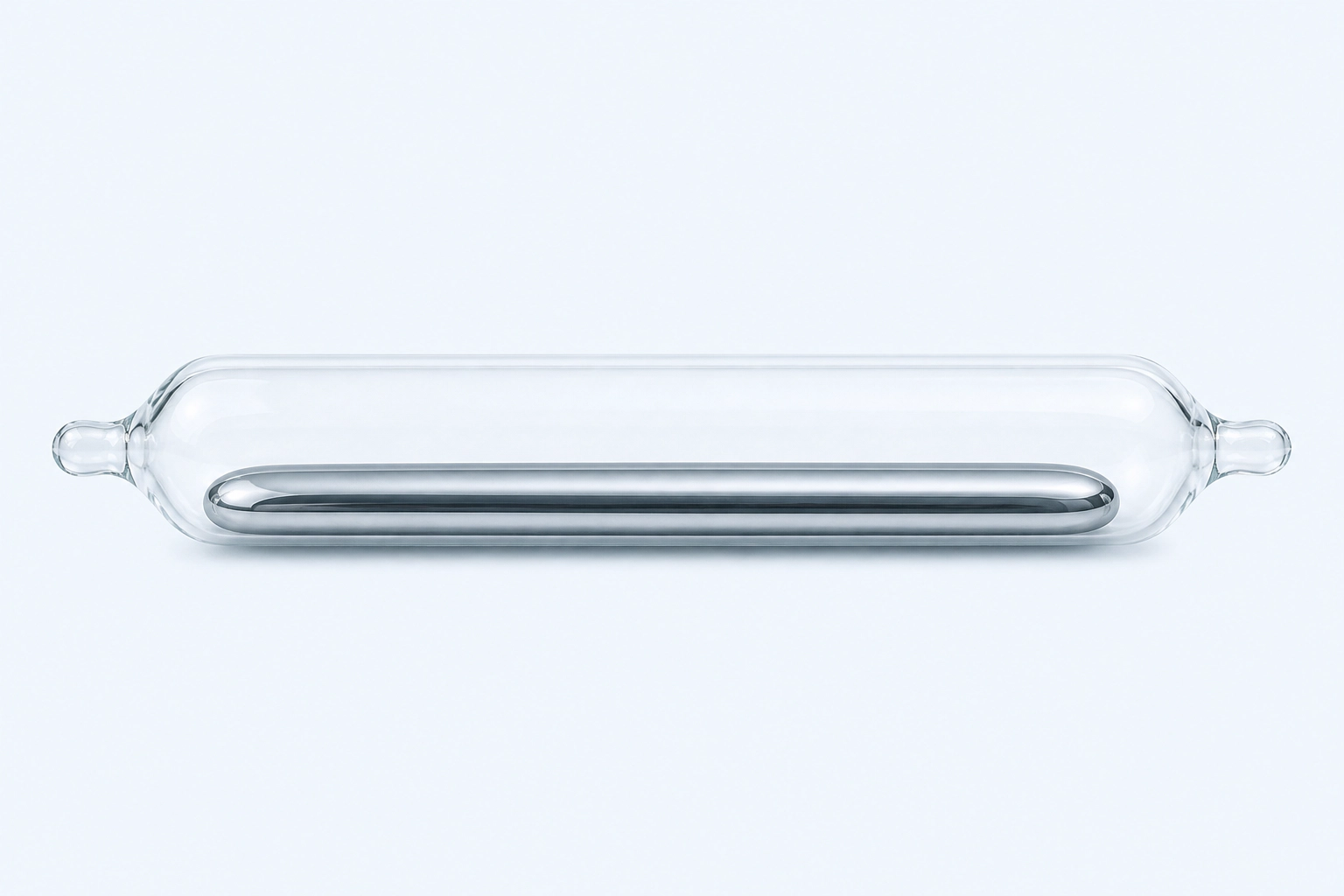
Elemental mercury (Hg), represented as one continuous silvery liquid body in a sealed ampoule. Containment does not certify purity and prevents this reconstruction from implying a spill or exposure; it is not analytical reference material or a photograph.
Scientific media record2 verified identifiers
- Subject
- Mercuryelement
- Identifiers
- Atomic number 80PubChem CID:23931
- Review
- Editorial review completeIdentifiers authority-verified · Accessibility validated · · mercury|mercury-technical-specimen-v1.webp
- Digital source
- Trained-algorithmic mediaCreated with a trained generative algorithm and reviewed by WikiBiome for subject identity, scientific framing, identifiers, provenance, and accessibility.
- Scientific basis
- Mercury — PubChem ElementMercury, PubChem CID 23931
- License
- CC BY-SA 4.0Created
Mercury is the most toxic heavy metal with no known biological function. Its organic form—methylmercury (methylmercury (MeHg))—is the primary concern for dietary exposure via fish consumption.
Mercury's toxicity centers on thiol group binding, glutathione depletion, and blood-brain barrier penetration, making it an especially potent neurotoxin.[1]Toxicity, Mechanism and Health Effects of Some Heavy MetalsMonisha Jaishankar, Tenzin Tseten, Naresh Anbalagan et al. · 2014Open reference 1 ↓
What sets mercury apart from other toxic metals in ecological terms is the Gut Microbiome's direct role in mercury speciation: intestinal bacteria both methylate inorganic mercury into the more toxic methylmercury form and demethylate methylmercury back to inorganic mercury, making the gut a critical metabolic interface for mercury toxicity.[2]Influence of Toxic Metal Exposure on the Gut Microbiota (Review)Federica Giambo, Sebastiano Italia, Michele Teodoro et al. · 2021Open reference 2 ↓
Evidence map63 cited passagesInspect provenance +
Mercury is the most toxic heavy metal with no known biological function. Its organic form—methylmercury (MeHg)—is the primary concern for dietary exposure via fish consumption. Mercury's toxicity centers on thiol group binding, glutathione depletion, and blood-brain barrier penetration, making it an especially potent neurotoxin. What sets mercury apart
Elemental mercury (Hg0): Liquid metal that produces vapor at room temperature. Hg vapor is lipophilic, readily absorbed through the lungs (80% absorption), crosses the blood-brain barrier, and deposits in brain tissue. Hg0 at 550 ug/m3 causes cognitive impairment and hippocampal damage in rats.
Inorganic mercury (Hg2+): Mercuric salts poorly absorbed through the GI tract (~7-15%) but highly nephrotoxic. Primary biomarker: urinary mercury.
Organic mercury (MeHg, EtHg): Methylmercury is 95-100% absorbed in the intestinal tract, the highest absorption efficiency of any metal form. MeHg readily crosses both the blood-brain barrier and the placental barrier.
All forms share a strong affinity for sulfhydryl/thiol groups (-SH), enabling binding to glutathione, cysteine residues, and metallothioneins. This thiol affinity is the unifying mechanism of mercury toxicity across organ systems.
Mercury has no known biological function in any living organism. It is purely toxic at all concentrations. However, multiple bacterial species have evolved mercury resistance mechanisms (merA reductase, merB organomercury lyase) as an adaptation to environmental mercury, and these resistance systems have deep evolutionary roots,.
Thiol binding and glutathione depletion. Hg binds to GSH and sulfhydryl groups on proteins, depleting the cell's primary antioxidant defense. Hg conjugates GSH, inhibits glutathione peroxidase, and disrupts the entire thiol-dependent antioxidant network,. In PCOS patients, serum mercury shows a strong negative correlation with GSH levels (P < 0.01), directly
ROS generation. Beyond GSH depletion, Hg directly stimulates reactive oxygen species production, creating a dual assault on cellular redox balance. heavy metals collectively impair carbohydrate, lipid, and amino acid metabolism, with ROS as the central mediator.
Enzyme inhibition. Hg inactivates enzymes through thiol binding, disrupting critical metabolic pathways. In the kidney, aquaporin mRNA is reduced, impairing water transport, and Na+/H+ exchangers and aquaporin-1 are inhibited in kidney tubules.
Neuroinflammation and demyelination. Hg triggers glial reactivity, increases TNF, IL-1, IL-6, and generates autoantibodies against neuronal proteins. MeHg downregulates myelin basic protein (MBP) expression, contributing to axonal demyelination in the CNS.
Cardiovascular damage. MeHg drives cardiovascular toxicity through ROS/lipid peroxidation, LDL oxidation, PLA2 activation, and inactivation of paraoxonase (PON), which reduces HDL's protective capacity. Perinatal MeHg exposure has been linked to hypertension onset in adolescence.
Hepatotoxicity. MeHg exposure alters bile acid metabolism and cholesterol pathways, with metabolomics revealing Hg-specific perturbations in the gut-liver axis.
Autoimmune induction. Mercury exposure induces lupus-like syndrome in autoimmune-prone mice, altering B cell receptor signaling via SYK phosphorylation, reducing Bank1 expression, and increasing NF-kB and TLR-9 activation.
Thyroid disruption. Mercury interferes with TSH production, inhibits thyroid peroxidase (TPO), and both MeHg and inorganic Hg compounds inhibit thyroglobulin (Tg) iodination—the critical step in thyroid hormone synthesis.
Fish consumption is the dominant source of MeHg exposure for the general population. Predatory species (tuna, swordfish, shark) bioaccumulate MeHg through the aquatic food chain. This creates the fish consumption paradox: fish is both the main MeHg source and provides neuroprotective omega-3 fatty acids and selenium, complicating risk assessment.
In Japanese children, MeHg accounted for approximately 90% of total Hg in diet samples where THg exceeded 1 ng/g, confirming fish as the overwhelmingly dominant dietary mercury source. Peak MeHg intake occurred at baby food stages 3 and 4 (9-17 months), reaching 346.6 ng/kg bw/day—substantially higher than formula milk (2.2 ng/kg bw/day) and exceeding ref
Canned fish (sardine, mackerel) is a significant exposure source in populations with high fish consumption. In Jamaican children, canned fish consumption showed a significant interaction with glutathione S-transferase genotype in predicting blood mercury: children with GSTP1 Ile/Ile genotype had 59% higher mean Hg with canned fish consumption, while those wi
Dental amalgam fillings contain approximately 50% mercury and release low levels of Hg vapor during chewing. Amalgam exposure increases Hg-resistant and antibiotic-resistant gut bacteria, establishing the oral cavity as a source of metal resistance gene dissemination to the intestinal microbiome,.
Occupational exposure occurs in artisanal gold mining, chloralkali plants, and instrument manufacturing. Environmental sources include coal combustion, volcanic emissions, and contaminated water systems. Korean adults have substantially higher blood mercury levels than US, Canadian, and European populations (3.11 vs 0.75 ug/L), reflecting dietary and environ
An estimated 8-10% of American women have mercury levels that could induce neurological disorders in children. Mercury crosses the placental membrane, causing spontaneous abortions, premature births, and congenital defects. Hair THg in Japanese children showed only weak correlation with dietary MeHg (r=0.170), suggesting that diet alone does not fully explai
Thimerosal (EtHg preservative in some vaccines) represents a medical exposure route. Higher EtHg was associated with lower scores on animal fluency and CERAD delayed recall in adults.
The gut microbiome is a critical metabolic interface for mercury. A core process in MeHg metabolism is the methylation/demethylation cycle carried out by intestinal bacteria: gut microbes can both methylate inorganic Hg into the more toxic MeHg form and demethylate MeHg back to inorganic Hg, altering mercury's speciation, toxicity, and bioavailability within
The gut microbiota serves as the first line of defense against heavy metal toxicity through bioaccumulation, binding, and enzymatic transformation of mercury, facilitating fecal excretion. Specific microbial mechanisms include metal transport proteins, sulfide production by sulfate-reducing bacteria, and biotransformation pathways. Approximately 60% of inges
Bacteroidetes enrichment: MeHg exposure increases Bacteroidetes at the phylum level and alters gut-brain related metabolites in animal models.
Showing 24 of 63 evidence-bearing passages. Every remaining citation is still indexed in the reference record below.
Contents
1. Chemical Properties and Forms2. Biological Roles3. Dietary and Environmental Sources4. Microbiome Interactions5. Nutritional Immunity6. Conditions Associated7. Interactions with Other Metals8. Biomarkers9. Key Studies10. Open Questions11. Cross-ReferencesChemical Properties and Forms#
Mercury exists in three toxicologically distinct forms, each with different absorption, distribution, and target organs. Elemental mercury (Hg0): Liquid metal that produces vapor at room temperature. mercury (Hg) vapor is lipophilic, readily absorbed through the lungs (80% absorption), crosses the blood-brain barrier, and deposits in brain tissue.
Hg0 at 550 ug/m3 causes cognitive impairment and hippocampal damage in rats.[3]Toxic Mechanisms of Five Heavy Metals: Mercury, Lead, Chromium, Cadmium, and ArsenicBalali-Mood M, Naseri K, Tahergorabi Z et al. · 2021Open reference 3 ↓
Inorganic mercury (mercury(II)): Mercuric salts poorly absorbed through the GI tract (~7-15%) but highly nephrotoxic. Primary biomarker: urinary mercury.[4]Molecular Mechanisms of Cellular Injury and Role of Toxic Heavy Metals in Chronic Kidney DiseaseManish Mishra, Larry Nichols, Aditi A. Dave et al. · 2022Open reference 4 ↓
Organic mercury (methylmercury, EtHg): Methylmercury is 95-100% absorbed in the intestinal tract, the highest absorption efficiency of any metal form. methylmercury readily crosses both the blood-brain barrier and the placental barrier.[1]Toxicity, Mechanism and Health Effects of Some Heavy MetalsMonisha Jaishankar, Tenzin Tseten, Naresh Anbalagan et al. · 2014Open reference 1 ↓
All forms share a strong affinity for sulfhydryl/thiol groups (-SH), enabling binding to glutathione, cysteine residues, and metallothioneins. This thiol affinity is the unifying mechanism of mercury toxicity across organ systems.[3]Toxic Mechanisms of Five Heavy Metals: Mercury, Lead, Chromium, Cadmium, and ArsenicBalali-Mood M, Naseri K, Tahergorabi Z et al. · 2021Open reference 3 ↓
Biological Roles#
Mercury has no known biological function in any living organism. It is purely toxic at all concentrations.[1]Toxicity, Mechanism and Health Effects of Some Heavy MetalsMonisha Jaishankar, Tenzin Tseten, Naresh Anbalagan et al. · 2014Open reference 1 ↓
However, multiple bacterial species have evolved mercury resistance mechanisms (merA reductase, merB organomercury lyase) as an adaptation to environmental mercury, and these resistance systems have deep evolutionary roots.[5]Baker-Austin 2006 — Co-selection of Antibiotic and Metal ResistanceBaker-Austin C, Wright MS, Stepanauskas R et al. · 2006Open reference 5 ↓[6]Diversity of metal and antibiotic resistance genes in Enterococcus spp. from the last century reflects multiple pollution and genetic exchange among phyla from overlapping ecosystemsRebelo A, Mourao J, Freitas AR et al. · 2021Open reference 6 ↓
Mechanism of Toxicity#
Mercury exerts toxicity through several converging pathways:
Thiol binding and glutathione depletion. mercury (Hg) binds to GSH and sulfhydryl groups on proteins, depleting the cell's primary antioxidant defense. mercury conjugates GSH, inhibits glutathione peroxidase, and disrupts the entire thiol-dependent antioxidant network.[3]Toxic Mechanisms of Five Heavy Metals: Mercury, Lead, Chromium, Cadmium, and ArsenicBalali-Mood M, Naseri K, Tahergorabi Z et al. · 2021Open reference 3 ↓[1]Toxicity, Mechanism and Health Effects of Some Heavy MetalsMonisha Jaishankar, Tenzin Tseten, Naresh Anbalagan et al. · 2014Open reference 1 ↓
In PCOS patients, serum mercury shows a strong negative correlation with GSH levels (P < 0.01), directly demonstrating this depletion mechanism in a clinical population.[7]Antioxidant Status in Relation to Heavy Metals Induced Oxidative Stress in Patients with Polycystic Ovarian Syndrome (PCOS)Manal Abudawood, Hajera Tabassum, Atheer H. Alanazi et al. · 2021Open reference 7 ↓
ROS generation. Beyond GSH depletion, mercury directly stimulates reactive oxygen species production, creating a dual assault on cellular redox balance.[3]Toxic Mechanisms of Five Heavy Metals: Mercury, Lead, Chromium, Cadmium, and ArsenicBalali-Mood M, Naseri K, Tahergorabi Z et al. · 2021Open reference 3 ↓ Heavy Metals collectively impair carbohydrate, lipid, and amino acid metabolism, with ROS as the central mediator.[8]Metabolomics: a promising tool for deciphering metabolic impairment in heavy metal toxicitiesAkash MSH, Yaqoob A, Rehman K et al. · 2023Open reference 8 ↓
Enzyme inhibition. mercury inactivates enzymes through thiol binding, disrupting critical metabolic pathways. In the kidney, aquaporin mRNA is reduced, impairing water transport, and Na+/H+ exchangers and aquaporin-1 are inhibited in kidney tubules.[4]Molecular Mechanisms of Cellular Injury and Role of Toxic Heavy Metals in Chronic Kidney DiseaseManish Mishra, Larry Nichols, Aditi A. Dave et al. · 2022Open reference 4 ↓
Neuroinflammation and demyelination. mercury triggers glial reactivity, increases TNF, IL-1, IL-6, and generates autoantibodies against neuronal proteins. methylmercury downregulates myelin basic protein (MBP) expression, contributing to axonal demyelination in the CNS.[9]Metal Profiles in Autism Spectrum Disorders: A Crosstalk between Toxic and Essential MetalsBlazewicz A, Grabrucker AM · 2023Open reference 9 ↓
Cardiovascular damage. methylmercury drives cardiovascular toxicity through ROS/lipid peroxidation, LDL oxidation, PLA2 activation, and inactivation of paraoxonase (PON), which reduces HDL's protective capacity. Perinatal methylmercury exposure has been linked to hypertension onset in adolescence.[10]Nucera 2024 — Non-essential heavy metal effects in cardiovascular diseases: overview of systematic reviewsNucera S, Serra M, Caminiti R et al. · 2024Open reference 10 ↓
Hepatotoxicity. methylmercury exposure alters bile acid metabolism and cholesterol pathways, with metabolomics revealing mercury-specific perturbations in the gut-liver axis.[8]Metabolomics: a promising tool for deciphering metabolic impairment in heavy metal toxicitiesAkash MSH, Yaqoob A, Rehman K et al. · 2023Open reference 8 ↓
Autoimmune induction. Mercury exposure induces lupus-like syndrome in autoimmune-prone mice, altering B cell receptor signaling via SYK phosphorylation, reducing Bank1 expression, and increasing NF-kB and TLR-9 activation.[11]Environmental Exposures and Autoimmune Diseases: Contribution of Gut MicrobiomeM. Firoze Khan, Hui Wang · 2020Open reference 11 ↓
Thyroid disruption. Mercury interferes with TSH production, inhibits thyroid peroxidase (TPO), and both methylmercury and inorganic mercury compounds inhibit thyroglobulin (Tg) iodination—the critical step in thyroid hormone synthesis.[12]Effects of Trace Elements on Endocrine Function and Pathogenesis of Thyroid Diseases — A Literature ReviewBrylinski L, Kostelecka K, Wolinski F et al. · 2025Open reference 12 ↓
Dietary and Environmental Sources#
Fish and Seafood (Primary Dietary Route)#
Fish consumption is the dominant source of methylmercury (MeHg) exposure for the general population. Predatory species (tuna, swordfish, shark) bioaccumulate methylmercury through the aquatic food chain.
This creates the fish consumption paradox: fish is both the main methylmercury source and provides neuroprotective omega-3 fatty acids and selenium, complicating risk assessment.[13]Heavy Metals Exposure and Alzheimer's Disease and Related DementiasBakulski KM, Seo YA, Hickman RC et al. · 2020Open reference 13 ↓
In Japanese children, methylmercury accounted for approximately 90% of total mercury in diet samples where THg exceeded 1 ng/g, confirming fish as the overwhelmingly dominant dietary mercury source.
Peak methylmercury intake occurred at baby food stages 3 and 4 (9-17 months), reaching 346.6 ng/kg bw/day—substantially higher than formula milk (2.2 ng/kg bw/day) and exceeding reference doses in several children.[14]Tatsuta 2024 — Dietary intake of methylmercury by 0-5 years children using the duplicate diet method in JapanNozomi Tatsuta, Kaname Asato, Miyuki Iwai-Shimada et al. · 2024Open reference 14 ↓
Canned fish (sardine, mackerel) is a significant exposure source in populations with high fish consumption.
In Jamaican children, canned fish consumption showed a significant interaction with glutathione S-transferase genotype in predicting blood mercury: children with GSTP1 Ile/Ile genotype had 59% higher mean mercury with canned fish consumption, while those with Val/Val genotype showed no effect—demonstrating that genetic polymorphisms in detoxification enzymes create differential susceptibility to the same dietary exposure.[15]Zaman 2023 — Factors associated with blood mercury and GST gene interactions in Jamaican childrenSheikh Farzana Zaman, Maureen Samms-Vaughan, Sepideh Saroukhani et al. · 2023Open reference 15 ↓
Dental Amalgam#
Dental amalgam fillings contain approximately 50% mercury and release low levels of mercury (Hg) vapor during chewing.
Amalgam exposure increases mercury-resistant and antibiotic-resistant gut bacteria, establishing the oral cavity as a source of metal resistance gene dissemination to the intestinal microbiome.[16]Effects of Heavy Metals on Gut Barrier Integrity and Gut MicrobiotaSweta Ghosh, Syam P. Nukavarpu, Venkatakrishna Rao Jala · 2024Open reference 16 ↓[5]Baker-Austin 2006 — Co-selection of Antibiotic and Metal ResistanceBaker-Austin C, Wright MS, Stepanauskas R et al. · 2006Open reference 5 ↓
Occupational and Environmental#
Occupational exposure occurs in artisanal gold mining, chloralkali plants, and instrument manufacturing. Environmental sources include coal combustion, volcanic emissions, and contaminated water systems.
Korean adults have substantially higher blood mercury levels than US, Canadian, and European populations (3.11 vs 0.75 ug/L), reflecting dietary and environmental differences.[17]A Comparative Study on the Paradoxical Relationship Between Heavy Metal Exposure and Kidney FunctionJee Hyun Rho, Seungho Lee, Jung-Yeon Kwon et al. · 2025Open reference 17 ↓
Infant and Prenatal Exposure#
An estimated 8-10% of American women have mercury levels that could induce neurological disorders in children.[1]Toxicity, Mechanism and Health Effects of Some Heavy MetalsMonisha Jaishankar, Tenzin Tseten, Naresh Anbalagan et al. · 2014Open reference 1 ↓ Mercury crosses the placental membrane, causing spontaneous abortions, premature births, and congenital defects.[18]Female Fertility and Environmental PollutionCanipari R, De Santis L, Cecconi S · 2020Open reference 18 ↓
Hair THg in Japanese children showed only weak correlation with dietary methylmercury (MeHg) (r=0.170), suggesting that diet alone does not fully explain body burden—prenatal loading and interindividual variation in GSH-mediated elimination likely contribute.[14]Tatsuta 2024 — Dietary intake of methylmercury by 0-5 years children using the duplicate diet method in JapanNozomi Tatsuta, Kaname Asato, Miyuki Iwai-Shimada et al. · 2024Open reference 14 ↓
Thimerosal (EtHg preservative in some vaccines) represents a medical exposure route. Higher EtHg was associated with lower scores on animal fluency and CERAD delayed recall in adults.[19]Exposure to heavy metals and neurocognitive function in adults: a systematic reviewAlthomali RH, Abbood MA, Saleh EAM et al. · 2024Open reference 19 ↓
Microbiome Interactions#
This section represents content that has no equivalent on Wikipedia: the bidirectional relationship between mercury and the gut microbiome, mercury's role as a selective pressure on microbial communities, and the co-selection of mercury resistance with antibiotic resistance.
Mercury Metabolism by Gut Microbiota#
The gut microbiome is a critical metabolic interface for mercury.
A core process in methylmercury (MeHg) metabolism is the methylation/demethylation cycle carried out by intestinal bacteria: gut microbes can both methylate inorganic mercury into the more toxic methylmercury form and demethylate methylmercury back to inorganic mercury, altering mercury's speciation, toxicity, and bioavailability within the intestinal lumen.[2]Influence of Toxic Metal Exposure on the Gut Microbiota (Review)Federica Giambo, Sebastiano Italia, Michele Teodoro et al. · 2021Open reference 2 ↓
The gut microbiota serves as the first line of defense against heavy metal toxicity through bioaccumulation, binding, and enzymatic transformation of mercury, facilitating fecal excretion. Specific microbial mechanisms include metal transport proteins, sulfide production by sulfate-reducing bacteria, and biotransformation pathways.[20]Gut Microbiota: A Target for Heavy Metal Toxicity and a Probiotic Protective StrategyHui Duan, Leilei Yu, Fengwei Tian et al. · 2020Open reference 20 ↓
Approximately 60% of ingested heavy metals are absorbed in the intestine, meaning the microbial community's capacity to sequester and transform metals before absorption is a major determinant of systemic toxicity.[21]Heavy Metal-Gut Microbiota Interactions: Probiotics Modulation and Biosensors DetectionLiliana Anchidin-Norocel, Oana C. Iatcu, Andrei Lobiuc et al. · 2025Open reference 21 ↓
Mercury-Induced Dysbiosis#
Mercury exposure consistently disrupts gut microbiota composition across multiple study models. Bacteroidetes enrichment: methylmercury (MeHg) exposure increases Bacteroidetes at the phylum level and alters gut-brain related metabolites in animal models.[20]Gut Microbiota: A Target for Heavy Metal Toxicity and a Probiotic Protective StrategyHui Duan, Leilei Yu, Fengwei Tian et al. · 2020Open reference 20 ↓
Pathogenic taxa selection: mercury/methylmercury exposure increases pathogenic Streptococcus and Enterococcus in the gut, while decreasing beneficial taxa.[22]Toxic and Essential Metals: Metabolic Interactions with the Gut Microbiota and Health ImplicationsQinheng Zhu, Boyan Chen, Fu Zhang et al. · 2024Open reference 22 ↓
Collinsella enrichment: A systematic review of 3,000+ subjects found that mercury, along with arsenic, lead, and cadmium, consistently enriched Collinsella as a cross-metal pathobiont marker, alongside enrichment of Desulfovibrio (a hydrogen sulfide producer that may further damage the intestinal barrier).[23]Rezazadegan et al. 2025 — Heavy Metals and Gut Microbiota: A Systematic ReviewFatemeh Rezazadegan, Maryam Mahmoudi, Seyed Mohammad Mousavi · 2025Open reference 23 ↓
Neurodevelopmental consequences: methylmercury-induced gut microbiota changes are linked to neurodevelopmental effects in rat offspring, establishing a mercury-gut-brain axis.[2]Influence of Toxic Metal Exposure on the Gut Microbiota (Review)Federica Giambo, Sebastiano Italia, Michele Teodoro et al. · 2021Open reference 2 ↓
The relationship is bidirectional: mercury alters microbiota composition and metabolic profiles, while the microbiota in turn modifies mercury absorption by acting as a physical barrier, modifying pH and oxidative balance, and expressing detoxification enzymes.[20]Gut Microbiota: A Target for Heavy Metal Toxicity and a Probiotic Protective StrategyHui Duan, Leilei Yu, Fengwei Tian et al. · 2020Open reference 20 ↓[22]Toxic and Essential Metals: Metabolic Interactions with the Gut Microbiota and Health ImplicationsQinheng Zhu, Boyan Chen, Fu Zhang et al. · 2024Open reference 22 ↓
Gut Barrier Disruption#
Mercury directly damages the intestinal epithelial barrier. mercury (Hg) downregulates claudin 1, occludin, ZO-1, and JAM1 in colon epithelial cells, increasing intestinal permeability and enabling bacterial translocation.[16]Effects of Heavy Metals on Gut Barrier Integrity and Gut MicrobiotaSweta Ghosh, Syam P. Nukavarpu, Venkatakrishna Rao Jala · 2024Open reference 16 ↓[21]Heavy Metal-Gut Microbiota Interactions: Probiotics Modulation and Biosensors DetectionLiliana Anchidin-Norocel, Oana C. Iatcu, Andrei Lobiuc et al. · 2025Open reference 21 ↓
This barrier disruption compounds the Dysbiosis: mercury-driven permeability allows LPS and other bacterial products to translocate systemically, driving neuroinflammation and immune activation through the gut-brain axis.[24]Ghasemian Sorboni 2023 — Comprehensive Review on Gut Microbiome in Neurological DisordersShokufeh Ghasemian Sorboni, Hanieh Shakeri Moghaddam, Reza Jafarzadeh-Esfehani et al. · 2023Open reference 24 ↓
Seven rodent studies confirm mercury causes intestinal barrier dysfunction, structural damage, gut Metal-Driven Inflammation, and microbiota dysbiosis.[25]Metal Dyshomeostasis as a Driver of Gut Pathology in Autism Spectrum DisordersO'Grady K, Grabrucker AM · 2025Open reference 25 ↓ Lactobacillus brevis 23017 protects against mercury-induced gut damage via MAPK and NF-kappaB pathway regulation.[16]Effects of Heavy Metals on Gut Barrier Integrity and Gut MicrobiotaSweta Ghosh, Syam P. Nukavarpu, Venkatakrishna Rao Jala · 2024Open reference 16 ↓
Mercury-Gut-Brain Metabolite Axis#
Mercury's disruption of the gut microbiome has downstream effects on neurologically active metabolites. In children, heavy metal load (including mercury) correlated with altered microbiome-associated catecholamine precursor metabolites (phenylalanine, tyrosine, L-dopa derivatives), accounting for 32% of variance in social behaviors.
Children with the lowest social behaviors had a sixfold increase in odds of high heavy metal loads.[26]Krajewski 2025 -- Heavy metals, noradrenaline/adrenaline ratio, and microbiome-associated hormone precursor metabolites: biomarkers for social behaviour, ADHD symptoms, and executive function in childrenKristin Krajewski · 2025Open reference 26 ↓
Mercury burden compounds SCFA depletion and impairs histone deacetylase inhibition, contributing to failure to upregulate metal-binding proteins and maintain intestinal barrier integrity.[24]Ghasemian Sorboni 2023 — Comprehensive Review on Gut Microbiome in Neurological DisordersShokufeh Ghasemian Sorboni, Hanieh Shakeri Moghaddam, Reza Jafarzadeh-Esfehani et al. · 2023Open reference 24 ↓
Mercury Resistance and Antibiotic Co-Selection#
Mercury is one of the most important metals in the co-selection of antibiotic resistance—a phenomenon with no equivalent coverage in standard mercury toxicology. Three distinct molecular mechanisms link mercury resistance to antibiotic resistance:
- cobalt (Co)-resistance: Mercury resistance genes and antibiotic resistance gene cassettes are physically linked on Tn21-like transposons and integrons. Integron In2 carries both mercury-resistance operons and aminoglycoside resistance genes (e.g., aadA1 for spectinomycin-streptomycin resistance).[5]Baker-Austin 2006 — Co-selection of Antibiotic and Metal ResistanceBaker-Austin C, Wright MS, Stepanauskas R et al. · 2006Open reference 5 ↓
- Cross-resistance: Shared efflux pump systems confer resistance to both mercury and antibiotics. Pseudomonas aeruginosa carries mercury resistance linked to multidrug resistance phenotypes.[27]Understanding the Development of Environmental Resistance Among Microbes: A ReviewSrivastava J, Chandra H, Singh N et al. · 2016Open reference 27 ↓
- cobalt-regulatory mechanisms: Mercury exposure transcriptionally activates resistance pathways that simultaneously confer antibiotic resistance, with mex and czc operons linking mercury efflux (merE) to imipenem resistance.[5]Baker-Austin 2006 — Co-selection of Antibiotic and Metal ResistanceBaker-Austin C, Wright MS, Stepanauskas R et al. · 2006Open reference 5 ↓
Enterococcus species carry the merA gene (encoding mercuric reductase) at 97% prevalence among mercury-resistant isolates, with six phylogenetic MerA variants identified across diverse ecological contexts (human, animal, food, aquatic). These mercury resistance genes co-occur with antibiotic resistance genes including vanA (vancomycin resistance) and erm(B) (macrolide resistance) on conjugative plasmids.
The temporal trend shows increasing co-selection of mercury tolerance and antibiotic resistance over 120 years, with acceleration since the 1990s.[6]Diversity of metal and antibiotic resistance genes in Enterococcus spp. from the last century reflects multiple pollution and genetic exchange among phyla from overlapping ecosystemsRebelo A, Mourao J, Freitas AR et al. · 2021Open reference 6 ↓
Critically, heavy metals are non-degradable, meaning mercury contamination represents a long-term, persistent selection pressure for antibiotic resistance that does not diminish over time—unlike antibiotic residues, which degrade.[5]Baker-Austin 2006 — Co-selection of Antibiotic and Metal ResistanceBaker-Austin C, Wright MS, Stepanauskas R et al. · 2006Open reference 5 ↓
Dental amalgam mercury has been directly linked to co-selection of antibiotic-resistant bacteria from both oral and intestinal communities.[5]Baker-Austin 2006 — Co-selection of Antibiotic and Metal ResistanceBaker-Austin C, Wright MS, Stepanauskas R et al. · 2006Open reference 5 ↓
Antifungal Properties#
In an intriguing counterpoint to its toxicity, mercury ions at 10 mM showed 100% susceptibility (i.e., 100% kill) of all 26 Candida strains isolated from HIV-positive patients. Mercury and cadmium ions inhibit the plasma membrane H+-ATPase of Candida, causing gradual loss of membrane potential.[28]Effect of Heavy Metal Ions on Candida Isolated from HIV Positive PatientsMonal M. Kukde, Silpi Basak, Deepak P. Selkar · 2019Open reference 28 ↓
This differential susceptibility—where bacteria evolve mercury resistance but fungi remain susceptible—highlights the ecological complexity of mercury in polymicrobial environments.
Nutritional Immunity#
Mercury is not a metal that the host deliberately sequesters through nutritional immunity (as iron and zinc are). Instead, the host's primary defense against mercury is glutathione conjugation and excretion, mediated by glutathione S-transferase (GST) enzymes.
Genetic variation in GST enzymes creates a wide spectrum of individual susceptibility to mercury. GSTT1, GSTM1, and GSTP1 polymorphisms modify the host's capacity to conjugate and eliminate mercury (Hg).
Null (DD) genotypes for GSTT1 and GSTM1 impair detoxification capacity, while GSTP1 Ile105Val (rs1695) modifies the gene-environment interaction: carriers of the Ile/Ile genotype who consume canned fish show 59% higher mean blood mercury, while Val/Val carriers show no elevation.[15]Zaman 2023 — Factors associated with blood mercury and GST gene interactions in Jamaican childrenSheikh Farzana Zaman, Maureen Samms-Vaughan, Sepideh Saroukhani et al. · 2023Open reference 15 ↓
Selenium-mercury antagonism is the most important protective interaction. Selenium reduces mercury toxicity by binding to mercury and facilitating biliary excretion.
In the thyroid—the organ with the highest selenium concentration in the body—selenium builds the deiodinases (DIO1, DIO2, DIO3) essential for T4 to T3 conversion and the glutathione peroxidases (GPx) protecting thyrocytes from oxidative damage.[29]McGregor Brock 2015 — The Role of Selenium in Thyroid Autoimmunity: A ReviewMcGregor Brock · 2015Open reference 29 ↓[12]Effects of Trace Elements on Endocrine Function and Pathogenesis of Thyroid Diseases — A Literature ReviewBrylinski L, Kostelecka K, Wolinski F et al. · 2025Open reference 12 ↓
Mercury's inhibition of TPO and Tg iodination thus compounds selenium depletion: mercury both directly impairs thyroid function and depletes the selenium needed for selenoprotein-dependent thyroid hormone metabolism.[12]Effects of Trace Elements on Endocrine Function and Pathogenesis of Thyroid Diseases — A Literature ReviewBrylinski L, Kostelecka K, Wolinski F et al. · 2025Open reference 12 ↓
Alpha-klotho has recently been identified as a mediator of mercury's nephrotoxic effects. Alpha-klotho mediates the mercury-CKD association with a 34.55% mediation proportion, and Mendelian randomization confirmed that higher alpha-klotho levels are causally associated with reduced CKD risk (OR 0.9842).
Proposed renoprotective functions include antioxidant enzyme regulation (SOD, CAT, GPX-4), TLR4 signaling suppression, NF-kappaB inhibition, and autophagy promotion.[30]The association between low-concentration heavy metal exposure and chronic kidney disease risk through alpha-klothoLiu S, Wang H, Cao Y et al. · 2025Open reference 30 ↓
Conditions Associated#
Neurodegenerative Disease#
Alzheimer's disease. mercury (Hg) increases amyloid-beta production and reduces its clearance. Mercury reduces neprilysin expression in SH-SY5Y neuronal cells, impairing the primary enzyme that degrades amyloid-beta.[31]Environmental pollutants as risk factors for neurodegenerative disorders: Alzheimer and Parkinson diseasesChin-Chan M, Navarro-Yepes J, Quintanilla-Vega B · 2015Open reference 31 ↓
Late-onset Alzheimer's disease (>95% of AD cases) results from cumulative metal burden interacting with genetic risk (APOE) and age-related vulnerability—declining blood-brain barrier integrity and impaired metal clearance amplify mercury neurotoxicity in aging brains.[32]Contributions of heavy metal exposure to late-onset Alzheimer's diseaseBakulski KM, et al. · 2025Open reference 32 ↓
However, the fish consumption paradox complicates epidemiological interpretation: fish as the main methylmercury source also provides neuroprotective omega-3s, and occupational mercury exposure studies show mixed results for dementia risk.[13]Heavy Metals Exposure and Alzheimer's Disease and Related DementiasBakulski KM, Seo YA, Hickman RC et al. · 2020Open reference 13 ↓[33]Exposure of metal toxicity in Alzheimer's disease: An extensive reviewIslam F, Shohag S, Akhter S et al. · 2022Open reference 33 ↓
Mercury crosses the BBB as vapor or methylmercury, accumulates in the cerebellum and cerebral cortex, disrupts glutamate transport, and impairs mitochondrial function.[34]Heavy metals in the diet: unraveling the molecular pathways linked to neurodegenerative disease riskGuevara-Ramirez P, Tamayo-Trujillo R, Cadena-Ullauri S et al. · 2024Open reference 34 ↓
Neurocognitive decline. Cadmium and mercury are the two metals most consistently associated with cognitive decline in adults across systematic reviews. Significant correlation exists between mercury exposure and deleterious neurocognitive outcomes, with elemental mercury exposure linked to significant reduction in short-term memory capacity in adults.
Notably, prenatal mercury exposure was NOT associated with lower cognitive scores in adulthood, suggesting a critical developmental window rather than cumulative lifetime dose.[19]Exposure to heavy metals and neurocognitive function in adults: a systematic reviewAlthomali RH, Abbood MA, Saleh EAM et al. · 2024Open reference 19 ↓
Autism Spectrum Disorder#
Mercury is elevated in blood, urine, hair, and teeth of ASD children.[9]Metal Profiles in Autism Spectrum Disorders: A Crosstalk between Toxic and Essential MetalsBlazewicz A, Grabrucker AM · 2023Open reference 9 ↓
In a pilot study of 136 Chinese children, mercury was significantly elevated in ASD hair samples, and trace element elevations co-varied with dysbiotic taxa enrichment, suggesting common selective pressure.[35]Zhai 2019 — Disturbance of Trace Elements and Gut Microbiota Profiles in AutismQixiao Zhai, Shi Cen, Jinchi Jiang et al. · 2019Open reference 35 ↓
Key pathomechanisms include Oxidative Stress via GSH inhibition, neuroinflammation through microglial activation, axonal demyelination via MBP downregulation, and competition with zinc for protein binding sites.[9]Metal Profiles in Autism Spectrum Disorders: A Crosstalk between Toxic and Essential MetalsBlazewicz A, Grabrucker AM · 2023Open reference 9 ↓[36]The Metallome as a Link Between the 'Omes' in Autism Spectrum DisordersJanelle E. Stanton, Sigita Malijauskaite, Kieran McGourty et al. · 2021Open reference 36 ↓
The metallome—the totality of metal ions in the body—connects the proteome, transcriptome, epigenome, microbiome, metabolome, and lipidome through metalloprotein function, and mercury's competition with zinc for thiol binding sites disrupts this entire network.[36]The Metallome as a Link Between the 'Omes' in Autism Spectrum DisordersJanelle E. Stanton, Sigita Malijauskaite, Kieran McGourty et al. · 2021Open reference 36 ↓
However, in a meta-analysis of two prospective pregnancy cohorts (EARLI and MARBLES, n=401), prenatal mercury did not show a consistent association with ASD at age 3, while cadmium did (OR 1.69)—suggesting that prenatal cadmium may be a stronger ASD risk factor than prenatal mercury at typical exposure levels.[37]Dou 2024 — Exposure to Heavy Metals in Utero and Autism Spectrum Disorder at Age 3: A Meta-Analysis of Two Longitudinal CohortsJohn F. Dou, Rebecca J. Schmidt, Heather E. Volk et al. · 2024Open reference 37 ↓
Chronic Kidney Disease#
Mercury disrupts mitochondrial membrane potential, triggers oxidative stress, and causes cytoskeletal alterations in proximal tubule cells. CKD patients with reduced renal mass are at heightened susceptibility to mercury (Hg) nephrotoxicity.[4]Molecular Mechanisms of Cellular Injury and Role of Toxic Heavy Metals in Chronic Kidney DiseaseManish Mishra, Larry Nichols, Aditi A. Dave et al. · 2022Open reference 4 ↓
In the general Korean population, increased mercury levels associate with decreased eGFR (the expected nephrotoxic pattern), but in environmentally vulnerable areas a paradoxical reversal appears: higher blood mercury reduced the OR of eGFR decline by 45.3%, likely reflecting reduced renal excretion as kidneys fail—blood levels drop not because exposure drops, but because the kidneys can no longer excrete mercury.[17]A Comparative Study on the Paradoxical Relationship Between Heavy Metal Exposure and Kidney FunctionJee Hyun Rho, Seungho Lee, Jung-Yeon Kwon et al. · 2025Open reference 17 ↓
Among 51 pollutants screened by machine learning, heavy metals (cadmium (Cd), thallium (Tl), lead (Pb), mercury) were the most impactful on CKD risk. Alpha-klotho mediates 34.55% of the mercury-CKD association.[30]The association between low-concentration heavy metal exposure and chronic kidney disease risk through alpha-klothoLiu S, Wang H, Cao Y et al. · 2025Open reference 30 ↓
In the CKD gut, mercury resistance phenotypes appear alongside lead and arsenic resistance in culturable bacteria, with co-resistance between metals and antibiotics detected—meaning mercury accumulation in the CKD gut may co-select for antibiotic-resistant pathogens.[38]Miranda 2022 — Metal(loid) and Antibiotic Resistance in CKD Gut BacteriaMiranda, Rojas, Geisse et al. · 2022Open reference 38 ↓
Cardiovascular Disease#
An overview of 8 systematic reviews covering 153 studies and 160,000+ participants confirms that mercury exposure independently increases risk of atherosclerosis, CAD, hypertension, myocardial infarction, and stroke.[10]Nucera 2024 — Non-essential heavy metal effects in cardiovascular diseases: overview of systematic reviewsNucera S, Serra M, Caminiti R et al. · 2024Open reference 10 ↓
methylmercury (MeHg) exposure in pregnancy associates with higher diastolic blood pressure in 2 of 6 reviewed studies, and perinatal mercury exposure is linked to hypertension onset in adolescence.[10]Nucera 2024 — Non-essential heavy metal effects in cardiovascular diseases: overview of systematic reviewsNucera S, Serra M, Caminiti R et al. · 2024Open reference 10 ↓
The cardiovascular mechanism involves PON-1 inactivation (reducing HDL's protective capacity against LDL oxidation), glutathione depletion, and lipid peroxidation.
All four non-essential toxic metals (cadmium (Cd), mercury, arsenic (As), lead (Pb)) converge on shared cardiovascular damage pathways: ROS/oxidative stress, endothelial dysfunction via NO reduction, LDL oxidation, and displacement of essential metals from physiologic binding sites.[10]Nucera 2024 — Non-essential heavy metal effects in cardiovascular diseases: overview of systematic reviewsNucera S, Serra M, Caminiti R et al. · 2024Open reference 10 ↓[39]Use of an Exposome Approach to Understand the Effects of Exposures From the Natural, Built, and Social Environments on Cardio-Vascular Disease Onset, Progression, and OutcomesPaul D. Juarez, Darryl B. Hood, Min-Ae Song et al. · 2020Open reference 39 ↓
Polycystic Ovary Syndrome#
Mercury is consistently elevated in PCOS patients across multiple studies. In a case-control study, PCOS patients had mercury (Hg) levels of 2.2 vs 1.3 ppb in controls (p < 0.001).[40]Are Heavy Metal Exposure and Trace Element Levels Related to Metabolic and Endocrine Problems in Polycystic Ovary Syndrome?Kirmizi DA, Baser E, Turksoy VA et al. · 2020Open reference 40 ↓
A prospective case-control study (n=106) found PCOS patients had mercury of 2.68 +/- 0.50 ppb vs controls, with a strong negative correlation between mercury and GSH (P < 0.01).[7]Antioxidant Status in Relation to Heavy Metals Induced Oxidative Stress in Patients with Polycystic Ovarian Syndrome (PCOS)Manal Abudawood, Hajera Tabassum, Atheer H. Alanazi et al. · 2021Open reference 7 ↓
A systematic review of 15 studies confirmed that women with PCOS have increased blood mercury alongside elevated antimony, cadmium, and lead, with decreased zinc.[41]Heavy Metals and Essential Elements in Association with Oxidative Stress in Women with Polycystic Ovary Syndrome -- A Systematic ReviewSmovrsnik T, Virant-Klun I, Pinter B · 2023Open reference 41 ↓
Mercury acts as an endocrine disruptor that can alter hormonal balance, menstrual cycle, ovulation, and fertility.
It interacts with sulfhydryl groups in the non-enzymatic antioxidant system (GSH), forming organometallic complexes, and disrupts insulin gene promoter activity and gonadotropin levels.[41]Heavy Metals and Essential Elements in Association with Oxidative Stress in Women with Polycystic Ovary Syndrome -- A Systematic ReviewSmovrsnik T, Virant-Klun I, Pinter B · 2023Open reference 41 ↓[7]Antioxidant Status in Relation to Heavy Metals Induced Oxidative Stress in Patients with Polycystic Ovarian Syndrome (PCOS)Manal Abudawood, Hajera Tabassum, Atheer H. Alanazi et al. · 2021Open reference 7 ↓
Thyroid Disease#
Mercury interferes with thyroid function at multiple levels: inhibition of TPO (blocking iodination of thyroglobulin), interference with TSH production, and inhibition of deiodinase enzymes needed for T4-to-T3 conversion.[12]Effects of Trace Elements on Endocrine Function and Pathogenesis of Thyroid Diseases — A Literature ReviewBrylinski L, Kostelecka K, Wolinski F et al. · 2025Open reference 12 ↓
Selenium plays a protective role against mercury's thyroid effects, and the antagonistic selenium (Se)-mercury (Hg) relationship has a demonstrated protective effect when mercury levels are elevated.[29]McGregor Brock 2015 — The Role of Selenium in Thyroid Autoimmunity: A ReviewMcGregor Brock · 2015Open reference 29 ↓
Reproductive Health#
Mercury crosses the placental membrane and is correlated with PCOS, endometriosis, dysmenorrhea, and amenorrhea.[18]Female Fertility and Environmental PollutionCanipari R, De Santis L, Cecconi S · 2020Open reference 18 ↓ Mercury disrupts normal sperm motility and activity.[42]The Effects of Toxic Heavy Metals Lead, Cadmium and Copper on the Epidemiology of Male and Female InfertilityAliasgar Manouchehri, Sarima Shokri, Mohadesh Pirhadi et al. · 2022Open reference 42 ↓
However, blood mercury was NOT significantly associated with female infertility in NHANES data,[43]Association between heavy metals exposure and infertility among American women aged 20-44 years: A cross-sectional analysis from 2013 to 2018 NHANES dataLin J, Lin X, Qiu J et al. · 2023Open reference 43 ↓ and serum total mercury showed no significant association with bacterial vaginosis risk in a study of 2,493 women (unlike lead and cadmium, which showed strong associations).[44]Feng 2025 — Heavy Metal Exposure and Bacterial VaginosisYu-Xue Feng, Ming-Zhi Tan, Hui-Han Qiu et al. · 2025Open reference 44 ↓
Depression#
In NHANES data analyzed by Bayesian Kernel Machine Regression, mercury showed a negative association with depressive symptoms in women—counter to neurotoxicity expectations. Lead and cadmium had stronger impacts on depression.
This negative mercury-depression association may reflect confounding from fish consumption (mercury source but also omega-3 source).[45]Ogundare 2024 — Association of Combined Effect of Metals Exposure and Behavioral Factors on Depressive Symptoms in WomenOlamide Ogundare, Emmanuel Obeng-Gyasi · 2024Open reference 45 ↓[46]Early pregnancy essential and non-essential metal mixtures and maternal antepartum and postpartum depressive symptomsRokoff LB, Cardenas A, Lin PI et al. · 2023Open reference 46 ↓
Arthritis#
Mercury showed negative/protective SHAP values for both general arthritis (-0.004) and RA specifically (-0.009) in a machine learning analysis of NHANES data, suggesting a potentially complex or confounded relationship—again possibly reflecting the fish consumption paradox.[47]Analyzing the impact of heavy metal exposure on osteoarthritis and rheumatoid arthritis: an approach based on interpretable machine learningFan W, Pi Z, Kong K et al. · 2024Open reference 47 ↓
Interactions with Other Metals#
Selenium: The most important protective interaction. selenium (Se) sequesters mercury (Hg) and facilitates biliary excretion, reducing toxicity. However, this depletes selenium available for selenoprotein synthesis (deiodinases, glutathione peroxidases), creating a secondary deficiency.
In the thyroid, selenium deficiency is a risk factor for both Hashimoto's thyroiditis and Graves' disease, meaning mercury-driven selenium depletion may increase autoimmune thyroid risk.[29]McGregor Brock 2015 — The Role of Selenium in Thyroid Autoimmunity: A ReviewMcGregor Brock · 2015Open reference 29 ↓[12]Effects of Trace Elements on Endocrine Function and Pathogenesis of Thyroid Diseases — A Literature ReviewBrylinski L, Kostelecka K, Wolinski F et al. · 2025Open reference 12 ↓
Zinc: mercury competes with zinc (Zn) for protein binding sites, contributing to functional zinc deficiency. Approximately 20% of dietary zinc is absorbed by intestinal bacteria, so mercury-driven dysbiosis may compound zinc bioavailability problems.[36]The Metallome as a Link Between the 'Omes' in Autism Spectrum DisordersJanelle E. Stanton, Sigita Malijauskaite, Kieran McGourty et al. · 2021Open reference 36 ↓
Lead and Cadmium: Frequently co-elevated in disease states (PCOS, ASD, CKD). All four metals (arsenic (As), cadmium (Cd), lead (Pb), mercury) show significant positive intercorrelations suggesting common environmental co-exposure patterns.[7]Antioxidant Status in Relation to Heavy Metals Induced Oxidative Stress in Patients with Polycystic Ovarian Syndrome (PCOS)Manal Abudawood, Hajera Tabassum, Atheer H. Alanazi et al. · 2021Open reference 7 ↓ Combined exposure likely produces additive or synergistic effects on ROS generation and GSH depletion.
Iron: Iron maintenance prevents/reduces cadmium uptake in women of fertile age. Mercury does not share this specific interaction but may compound iron-mediated oxidative stress in inflamed tissues.[18]Female Fertility and Environmental PollutionCanipari R, De Santis L, Cecconi S · 2020Open reference 18 ↓
Calcium: mercury, like lead, can interfere with calcium-dependent processes. In the cardiovascular system, displacement of calcium(II) (Ca2+) from physiologic binding sites is one of the converging mechanisms by which toxic metals drive endothelial damage.[10]Nucera 2024 — Non-essential heavy metal effects in cardiovascular diseases: overview of systematic reviewsNucera S, Serra M, Caminiti R et al. · 2024Open reference 10 ↓
Biomarkers#
| Matrix | What It Reflects | Notes |
|---|---|---|
| Blood/whole blood mercury (Hg) | Recent methylmercury exposure | Primarily reflects organic mercury from fish; Korean adults average 3.11 ug/L vs US 0.75 ug/L[17]A Comparative Study on the Paradoxical Relationship Between Heavy Metal Exposure and Kidney FunctionJee Hyun Rho, Seungho Lee, Jung-Yeon Kwon et al. · 2025Open reference 17 ↓ |
| Urinary mercury | Inorganic mercury exposure | Reflects inorganic mercury and occupational/dental amalgam exposure; part of NHANES biomonitoring[30]The association between low-concentration heavy metal exposure and chronic kidney disease risk through alpha-klothoLiu S, Wang H, Cao Y et al. · 2025Open reference 30 ↓ |
| Hair mercury | Medium-term methylmercury exposure (3-6 months) | Commonly used in epidemiological studies; provides integrated exposure measurement. Hair THg geometric mean 1.05 ppm in Japanese children[14]Tatsuta 2024 — Dietary intake of methylmercury by 0-5 years children using the duplicate diet method in JapanNozomi Tatsuta, Kaname Asato, Miyuki Iwai-Shimada et al. · 2024Open reference 14 ↓ |
| Toenail mercury | Longer-term exposure (6-12 months) | Used in Sister Study and other cohorts |
| Cord blood | Prenatal methylmercury exposure | Reflects placental transfer; critical for neurodevelopmental risk assessment |
| Deciduous teeth | Cumulative in-utero and early-life exposure | ASD children show approximately 2-fold higher mercury than neurotypical controls[9]Metal Profiles in Autism Spectrum Disorders: A Crosstalk between Toxic and Essential MetalsBlazewicz A, Grabrucker AM · 2023Open reference 9 ↓ |
Key Studies#
| Source | Evidence Level | Key Mercury Finding |
|---|---|---|
| [10]Nucera 2024 — Non-essential heavy metal effects in cardiovascular diseases: overview of systematic reviewsNucera S, Serra M, Caminiti R et al. · 2024Open reference 10 ↓ | Systematic review | 8 SRs, 160,000+ subjects: mercury (Hg) independently increases CVD risk; PON-1 inactivation mechanism |
| [23]Rezazadegan et al. 2025 — Heavy Metals and Gut Microbiota: A Systematic ReviewFatemeh Rezazadegan, Maryam Mahmoudi, Seyed Mohammad Mousavi · 2025Open reference 23 ↓ | Systematic review | 3,000+ subjects: mercury disrupts gut microbiota; Collinsella and Desulfovibrio enriched |
| [41]Heavy Metals and Essential Elements in Association with Oxidative Stress in Women with Polycystic Ovary Syndrome -- A Systematic ReviewSmovrsnik T, Virant-Klun I, Pinter B · 2023Open reference 41 ↓ | Systematic review | 15 studies: mercury consistently elevated in PCOS with depleted zinc and antioxidants |
| [19]Exposure to heavy metals and neurocognitive function in adults: a systematic reviewAlthomali RH, Abbood MA, Saleh EAM et al. · 2024Open reference 19 ↓ | Systematic review | mercury and cadmium (Cd) most consistently associated with cognitive decline in adults |
| [5]Baker-Austin 2006 — Co-selection of Antibiotic and Metal ResistanceBaker-Austin C, Wright MS, Stepanauskas R et al. · 2006Open reference 5 ↓ | Expert opinion (seminal) | Established framework for mercury-antibiotic co-resistance via Tn21, integrons, efflux pumps |
| [20]Gut Microbiota: A Target for Heavy Metal Toxicity and a Probiotic Protective StrategyHui Duan, Leilei Yu, Fengwei Tian et al. · 2020Open reference 20 ↓ | Animal model (review) | Bidirectional mercury-microbiota interaction; Bacteroidetes enrichment; probiotic protective strategies |
| [35]Zhai 2019 — Disturbance of Trace Elements and Gut Microbiota Profiles in AutismQixiao Zhai, Shi Cen, Jinchi Jiang et al. · 2019Open reference 35 ↓ | Cross-sectional | mercury elevated in ASD hair; trace elements co-vary with dysbiotic taxa |
| [7]Antioxidant Status in Relation to Heavy Metals Induced Oxidative Stress in Patients with Polycystic Ovarian Syndrome (PCOS)Manal Abudawood, Hajera Tabassum, Atheer H. Alanazi et al. · 2021Open reference 7 ↓ | Case-control | mercury 2.68 ppb in PCOS vs controls; negative correlation with GSH (P<0.01) |
| [14]Tatsuta 2024 — Dietary intake of methylmercury by 0-5 years children using the duplicate diet method in JapanNozomi Tatsuta, Kaname Asato, Miyuki Iwai-Shimada et al. · 2024Open reference 14 ↓ | Cross-sectional | Peak methylmercury intake at 9-17 months in Japanese children; methylmercury ~90% of dietary mercury |
Open Questions#
Unresolved questions identified by the current evidence record.
01Gut microbial methylation balance: Which gut bacterial species drive the methylation vs. demethylation balance of mercury in the intestinal lumen?+
Shifting this balance could alter systemic methylmercury (MeHg) exposure without changing dietary intake.
02Fish consumption paradox: How can methylmercury (MeHg) neurotoxicity be disentangled from omega-3 neuroprotection in epidemiological studies?+
Mercury shows negative associations with depression and arthritis, likely confounded by fish-derived omega-3s.
03Dental amalgam and gut resistome: What is the quantitative contribution of dental amalgam to the gut microbiome's mercury resistance gene pool, and does amalgam removal reduce co-selected antibiotic resistance?+
The current WikiBiome record identifies this as an unresolved evidence gap.
04Selenium-mercury threshold: At what selenium (Se):mercury molar ratio does selenium's protective effect against mercury neurotoxicity saturate, and does fish-derived selenium adequately protect against fish-derived methylmercury (MeHg)?+
The current WikiBiome record identifies this as an unresolved evidence gap.
05Alpha-klotho as biomarker: Can alpha-klotho levels predict individual susceptibility to mercury-driven nephrotoxicity before GFR decline becomes clinically apparent?+
The current WikiBiome record identifies this as an unresolved evidence gap.
06Dose-response at low levels: At typical dietary exposure levels in developed countries, does methylmercury (MeHg) meaningfully contribute to neurodegeneration risk, or is the threshold higher than current population exposures?+
The current WikiBiome record identifies this as an unresolved evidence gap.
07Speciation in vivo: Chemical speciation of mercury (Hg) in tissues and biofluids is critically understudied; the oxidation state and molecular form likely determine bioavailability and toxicity.+
The current WikiBiome record identifies this as an unresolved evidence gap.
08GST pharmacogenomics: Can GST genotyping (GSTT1, GSTM1, GSTP1) stratify populations into high- and low-risk groups for dietary mercury exposure, enabling personalized fish consumption guidance?+
The current WikiBiome record identifies this as an unresolved evidence gap.
Cross-References#
Lead—co-reviewed neurotoxin; shared ASD and AD associations; both compete with zinc; co-elevated in PCOS and CKD. Cadmium—frequently co-elevated in disease states; both associated with neurocognitive decline; shared cardiovascular and reproductive toxicity.
Zinc—mercury (Hg) competes with zinc (Zn) for binding sites; functional zinc deficiency proposed as shared mechanism in ASD; 20% of dietary zinc absorbed by gut bacteria.
Selenium—key protective interaction; selenium (Se) sequesters mercury; selenium depletion impairs thyroid selenoprotein function. Copper—co-measured in PCOS and metallomics studies; copper (Cu)/zinc ratio elevated in ASD. Arsenic—co-reviewed toxic metal sharing kidney and neurological targets; co-elevated across disease states.
Nickel—both measured in PCOS and IBD studies; both drive co-selection of antibiotic resistance. Iron—mercury compounds iron-mediated oxidative stress; iron status modifies cadmium absorption. oxidative stress—thiol depletion and ROS as central mechanisms of mercury toxicity.
Glutathione (GSH)—mercury depletes GSH by binding thiol groups; GST polymorphisms modify elimination capacity. Antimicrobial Resistance—mercury resistance genes (merA, merR) physically linked to antibiotic resistance on Tn21 transposons. Enterococcus—merA at 97% prevalence; century-long temporal trend of increasing mercury-antibiotic co-resistance.
Co-Selection—mercury as a non-degradable, persistent driver of antibiotic resistance gene maintenance. Gut-Metal-Microbiome Interactions—bidirectional mercury-microbiota interaction; methylation/demethylation cycle; barrier disruption. Neurodegeneration and Metals—methylmercury crosses BBB; accumulates in CNS; implicated in AD and cognitive decline.
Cardiovascular Disease—PON-1 inactivation, LDL oxidation, endothelial dysfunction. Heavy Metals—mercury is the prototypical purely toxic heavy metal with no biological function. Biomarkers—blood, hair, urine, teeth, and cord blood mercury as exposure biomarkers across time windows.
Metal Chelation Therapy—DMSA, DMPS, and BAL used for mercury poisoning. Alpha-Klotho—mediates 34.55% of the mercury-CKD association; renoprotective via NF-kB suppression.
References 53
Numbered by first appearance in the article, then reconciled with its declared source list.
- 1
★Monisha Jaishankar, Tenzin Tseten, Naresh Anbalagan et al. (2014). Toxicity, Mechanism and Health Effects of Some Heavy Metals. Interdisciplinary Toxicology.
- 2
★Federica Giambo, Sebastiano Italia, Michele Teodoro et al. (2021). Influence of Toxic Metal Exposure on the Gut Microbiota (Review). World Academy of Sciences Journal.
- 3
★Balali-Mood M, Naseri K, Tahergorabi Z et al. (2021). Toxic Mechanisms of Five Heavy Metals: Mercury, Lead, Chromium, Cadmium, and Arsenic. Frontiers in Pharmacology.
- 4
★Manish Mishra, Larry Nichols, Aditi A. Dave et al. (2022). Molecular Mechanisms of Cellular Injury and Role of Toxic Heavy Metals in Chronic Kidney Disease. International Journal of Molecular Sciences.
- 5
Baker-Austin C, Wright MS, Stepanauskas R et al. (2006). Baker-Austin 2006 — Co-selection of Antibiotic and Metal Resistance. Trends in Microbiology.
- 6
Rebelo A, Mourao J, Freitas AR et al. (2021). Diversity of metal and antibiotic resistance genes in Enterococcus spp. from the last century reflects multiple pollution and genetic exchange among phyla from overlapping ecosystems. Science of the Total Environment.
- 7
Manal Abudawood, Hajera Tabassum, Atheer H. Alanazi et al. (2021). Antioxidant Status in Relation to Heavy Metals Induced Oxidative Stress in Patients with Polycystic Ovarian Syndrome (PCOS). Scientific Reports.
- 8
Akash MSH, Yaqoob A, Rehman K et al. (2023). Metabolomics: a promising tool for deciphering metabolic impairment in heavy metal toxicities. Frontiers in Molecular Biosciences.
- 9
Blazewicz A, Grabrucker AM (2023). Metal Profiles in Autism Spectrum Disorders: A Crosstalk between Toxic and Essential Metals. International Journal of Molecular Sciences.
- 10
Nucera S, Serra M, Caminiti R et al. (2024). Nucera 2024 — Non-essential heavy metal effects in cardiovascular diseases: overview of systematic reviews. Frontiers in Cardiovascular Medicine.
- 11
M. Firoze Khan, Hui Wang (2020). Environmental Exposures and Autoimmune Diseases: Contribution of Gut Microbiome. Frontiers in Immunology.
- 12
★Brylinski L, Kostelecka K, Wolinski F et al. (2025). Effects of Trace Elements on Endocrine Function and Pathogenesis of Thyroid Diseases — A Literature Review. Nutrients.
- 13
★Bakulski KM, Seo YA, Hickman RC et al. (2020). Heavy Metals Exposure and Alzheimer's Disease and Related Dementias. Journal of Alzheimer's Disease.
- 14
Nozomi Tatsuta, Kaname Asato, Miyuki Iwai-Shimada et al. (2024). Tatsuta 2024 — Dietary intake of methylmercury by 0-5 years children using the duplicate diet method in Japan. Environmental Health and Preventive Medicine.
- 15
Sheikh Farzana Zaman, Maureen Samms-Vaughan, Sepideh Saroukhani et al. (2023). Zaman 2023 — Factors associated with blood mercury and GST gene interactions in Jamaican children. BMC Pediatrics.
- 16
★Sweta Ghosh, Syam P. Nukavarpu, Venkatakrishna Rao Jala (2024). Effects of Heavy Metals on Gut Barrier Integrity and Gut Microbiota. Microbiota and Host.
- 17
Jee Hyun Rho, Seungho Lee, Jung-Yeon Kwon et al. (2025). A Comparative Study on the Paradoxical Relationship Between Heavy Metal Exposure and Kidney Function. Diagnostics.
- 18
Canipari R, De Santis L, Cecconi S (2020). Female Fertility and Environmental Pollution. International Journal of Environmental Research and Public Health.
- 19
Althomali RH, Abbood MA, Saleh EAM et al. (2024). Exposure to heavy metals and neurocognitive function in adults: a systematic review. Environmental Sciences Europe.
- 20
★Hui Duan, Leilei Yu, Fengwei Tian et al. (2020). Gut Microbiota: A Target for Heavy Metal Toxicity and a Probiotic Protective Strategy. Science of the Total Environment.
- 21
★Liliana Anchidin-Norocel, Oana C. Iatcu, Andrei Lobiuc et al. (2025). Heavy Metal-Gut Microbiota Interactions: Probiotics Modulation and Biosensors Detection. Biosensors.
- 22
★Qinheng Zhu, Boyan Chen, Fu Zhang et al. (2024). Toxic and Essential Metals: Metabolic Interactions with the Gut Microbiota and Health Implications. Frontiers in Nutrition.
- 23
Fatemeh Rezazadegan, Maryam Mahmoudi, Seyed Mohammad Mousavi (2025). Rezazadegan et al. 2025 — Heavy Metals and Gut Microbiota: A Systematic Review. Journal of Health, Population and Nutrition.
- 24
Shokufeh Ghasemian Sorboni, Hanieh Shakeri Moghaddam, Reza Jafarzadeh-Esfehani et al. (2023). Ghasemian Sorboni 2023 — Comprehensive Review on Gut Microbiome in Neurological Disorders. Unknown.
- 25
★O'Grady K, Grabrucker AM (2025). Metal Dyshomeostasis as a Driver of Gut Pathology in Autism Spectrum Disorders. Journal of Neurochemistry.
- 26
Kristin Krajewski (2025). Krajewski 2025 -- Heavy metals, noradrenaline/adrenaline ratio, and microbiome-associated hormone precursor metabolites: biomarkers for social behaviour, ADHD symptoms, and executive function in children. Scientific Reports.
- 27
Srivastava J, Chandra H, Singh N et al. (2016). Understanding the Development of Environmental Resistance Among Microbes: A Review. Clean - Soil, Air, Water.
- 28
Monal M. Kukde, Silpi Basak, Deepak P. Selkar (2019). Effect of Heavy Metal Ions on Candida Isolated from HIV Positive Patients. Journal of Clinical and Diagnostic Research.
- 29
McGregor Brock (2015). McGregor Brock 2015 — The Role of Selenium in Thyroid Autoimmunity: A Review. Journal of Restorative Medicine.
- 30
Liu S, Wang H, Cao Y et al. (2025). The association between low-concentration heavy metal exposure and chronic kidney disease risk through alpha-klotho. Scientific Reports.
- 31
★Chin-Chan M, Navarro-Yepes J, Quintanilla-Vega B (2015). Environmental pollutants as risk factors for neurodegenerative disorders: Alzheimer and Parkinson diseases. Frontiers in Cellular Neuroscience.
- 32
Bakulski KM, et al. (2025). Contributions of heavy metal exposure to late-onset Alzheimer's disease. (Behavioural Brain Research / related journal).
- 33
Islam F, Shohag S, Akhter S et al. (2022). Exposure of metal toxicity in Alzheimer's disease: An extensive review. Frontiers in Pharmacology.
- 34
★Guevara-Ramirez P, Tamayo-Trujillo R, Cadena-Ullauri S et al. (2024). Heavy metals in the diet: unraveling the molecular pathways linked to neurodegenerative disease risk. Food and Agricultural Immunology.
- 35
Qixiao Zhai, Shi Cen, Jinchi Jiang et al. (2019). Zhai 2019 — Disturbance of Trace Elements and Gut Microbiota Profiles in Autism. Environmental Research.
- 36
Janelle E. Stanton, Sigita Malijauskaite, Kieran McGourty et al. (2021). The Metallome as a Link Between the 'Omes' in Autism Spectrum Disorders. Frontiers in Molecular Neuroscience.
- 37
John F. Dou, Rebecca J. Schmidt, Heather E. Volk et al. (2024). Dou 2024 — Exposure to Heavy Metals in Utero and Autism Spectrum Disorder at Age 3: A Meta-Analysis of Two Longitudinal Cohorts. Environmental Health.
- 38
Miranda, Rojas, Geisse et al. (2022). Miranda 2022 — Metal(loid) and Antibiotic Resistance in CKD Gut Bacteria. Biological Research.
- 39
Paul D. Juarez, Darryl B. Hood, Min-Ae Song et al. (2020). Use of an Exposome Approach to Understand the Effects of Exposures From the Natural, Built, and Social Environments on Cardio-Vascular Disease Onset, Progression, and Outcomes. Frontiers in Public Health.
- 40
★Kirmizi DA, Baser E, Turksoy VA et al. (2020). Are Heavy Metal Exposure and Trace Element Levels Related to Metabolic and Endocrine Problems in Polycystic Ovary Syndrome?. Biological Trace Element Research.
- 41
Smovrsnik T, Virant-Klun I, Pinter B (2023). Heavy Metals and Essential Elements in Association with Oxidative Stress in Women with Polycystic Ovary Syndrome -- A Systematic Review. Antioxidants.
- 42
Aliasgar Manouchehri, Sarima Shokri, Mohadesh Pirhadi et al. (2022). The Effects of Toxic Heavy Metals Lead, Cadmium and Copper on the Epidemiology of Male and Female Infertility. JBRA Assisted Reproduction.
- 43
Lin J, Lin X, Qiu J et al. (2023). Association between heavy metals exposure and infertility among American women aged 20-44 years: A cross-sectional analysis from 2013 to 2018 NHANES data. Frontiers in Public Health.
- 44
Yu-Xue Feng, Ming-Zhi Tan, Hui-Han Qiu et al. (2025). Feng 2025 — Heavy Metal Exposure and Bacterial Vaginosis. PLOS ONE.
- 45
Olamide Ogundare, Emmanuel Obeng-Gyasi (2024). Ogundare 2024 — Association of Combined Effect of Metals Exposure and Behavioral Factors on Depressive Symptoms in Women. Toxics.
- 46
Rokoff LB, Cardenas A, Lin PI et al. (2023). Early pregnancy essential and non-essential metal mixtures and maternal antepartum and postpartum depressive symptoms. Neurotoxicology.
- 47
Fan W, Pi Z, Kong K et al. (2024). Analyzing the impact of heavy metal exposure on osteoarthritis and rheumatoid arthritis: an approach based on interpretable machine learning. Frontiers in Nutrition.
- 48
Jessica Briffa, Emmanuel Sinagra, Renald Blundell (2020). Heavy Metal Pollution in the Environment and Their Toxicological Effects on Humans. Heliyon.
- 49
Unknown - file inaccessible due to encoding. Evaluation of the Risk from Potentially Toxic Elements (PTEs) in Italy's Most Consumed Processed Fish Products. Unknown.
- 50
Smovrsnik T, Pinter B, Horvat M et al. (2025). Association of Trace Elements with Polycystic Ovary Syndrome in Women -- A Case-Control Study. Metabolites.
- 51
Xing Yan, Jun Qiu, Ruiwen Huang et al. (2025). Yan 2025 — Association Between Infants' Serum Levels of 26 Metals and Gut Microbiota: A Hospital-Based Cross-Sectional Study in China. Frontiers in Microbiology.
- 52
GB Rogers, DJ Keating, RL Young et al. (2016). Rogers 2016 -- From gut dysbiosis to altered brain function and mental illness: mechanisms and pathways. Molecular Psychiatry.
- 53
Du N, Du M, Punshon T et al. (2025). Heavy metal exposures in aerodigestive clinic cohort of infants with reflux or dysphagia. Scientific Reports.
Article network
Mentioned here 18
Pages linking here 48
Connect the evidence
Publicly readable discussion by ORCID-authenticated researchers. Questions, interpretation, methods, corrections, and new evidence stay attached to this record.
No discussion yet. Start with a precise question or a source-backed observation.
Activity and accepted changes
Accepted researcher context, editorial status, public discussion, and upstream Git revisions are shown together. Pending, declined, and withdrawn proposals remain private.
- published revision
Strengthen TLR4 and link high-leverage contexts
Karen Pendergrass · +1 −1
Inspect exact Git diff ↗ - published revision
Add reviewed neuroinflammation coverage batch
Karen Pendergrass · +1 −1
Inspect exact Git diff ↗ - published revision
Backfill heavy metals concept links
Karen Pendergrass · +2 −2
Inspect exact Git diff ↗ - published revision
Backfill oxidative stress concept links
Karen Pendergrass · +2 −2
Inspect exact Git diff ↗ - published revision
Backfill gut microbiome concept links
Karen Pendergrass · +1 −1
Inspect exact Git diff ↗ - published revision
Backfill inflammation concept links
Karen Pendergrass · +1 −1
Inspect exact Git diff ↗ - published revision
Complete corpus-wide Dysbiosis linking
Karen Pendergrass · +1 −1
Inspect exact Git diff ↗ - published revision
massive wiki expansion: 149 stubs fixed, 100+ new pages, Rule 15 scan, keystone papers
WikiBiome Deploy Bot · +56 −56
Inspect exact Git diff ↗ - published revision
deepen 7 metal entity pages: iron, zinc, cadmium, lead, mercury, nickel, arsenic
WikiBiome Deploy Bot · +203 −87
Inspect exact Git diff ↗ - published revision
Deepen metal/concept entities + 8 new sources for T1D/schizophrenia
WikiBiome Deploy Bot · +2 −2
Inspect exact Git diff ↗ - published revision
wiki: bulk entity upgrades, new article pages, and site regeneration
WikiBiome Deploy Bot · +6 −0
Inspect exact Git diff ↗ - published revision
WikiBiome v2 migration: signature pages + safety fixes + gap analysis
WikiBiome Deploy Bot · +6 −0
Inspect exact Git diff ↗ - published revision
WikiBiome update — integrity fixes, metallomic diet pages, cross-condition analyses
WikiBiome Deploy Bot · +3 −1
Inspect exact Git diff ↗ - published revision
WikiBiome v7 — interactive microbiome metallomics encyclopedia
Karen Pendergrass · +125 −0
Inspect exact Git diff ↗