
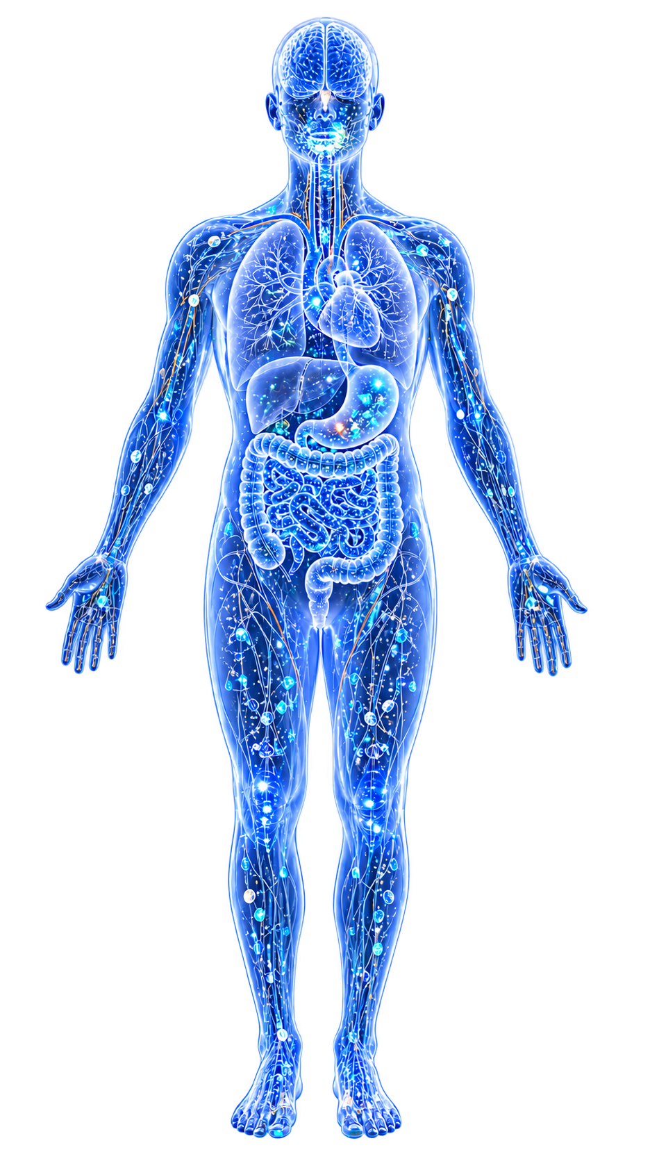
An umbrella concept encompassing the metallomic dimensions of Alzheimer's disease, Parkinson's disease, and other neurodegenerative conditions. The brain is uniquely vulnerable to metal toxicity: it has high metabolic demand, limited regenerative capacity, region-specific metal accumulation, and a blood-brain barrier (BBB) that metals can both bypass and damage.
Ferroptosis has emerged as the convergent cell death mechanism linking metal dyshomeostasis to neuronal loss across multiple diseases.
Evidence map13 cited passagesInspect provenance +
Substantia nigra: iron accumulation is the hallmark of PD; neuromelanin normally sequesters Fe but capacity can be exceeded.
Hippocampus and cortex: iron and zinc accumulate in AD; Cu is paradoxically depleted.
Amyloid plaques: enriched in Zn and Cu, which bind amyloid-beta directly and promote aggregation.
Post-mortem brain metallomics reveals widespread Cu decreases across multiple brain regions in AD, DLB, and PDD.
Lead and cadmium directly damage the BBB, increasing permeability to metals, toxins, and pathogens.
ferroptosis—iron-dependent lipid peroxidation leading to cell death—is the point of convergence for metal-driven neurodegeneration:
Neuromelanin iron-binding capacity modulates vulnerability: MC1R variants (red hair phenotype) may increase PD risk by shifting neuromelanin toward pheomelanin with weaker Fe chelation.
Iron chelation (deferiprone) shows some benefit in PD and AD trials.
All neurotoxic metals converge on overlapping pathways:
Neuroinflammation—metal-activated microglia via nf kappa b and NLRP3.
Epigenetic modifications—early-life Pb exposure produces latent AD-related gene expression changes decades later.
Alpha-synuclein aggregation may begin in the enteric nervous system and propagate to the brain via the vagus nerve (Braak hypothesis).
Aluminum accumulates in brain tissue in AD and is a documented neurotoxin, but its causal role remains debated. Al's neurotoxicity mechanisms include oxidative stress, inflammatory cytokine induction, and interference with iron homeostasis. The difficulty of measuring Al exposure accurately confounds epidemiological studies.
Contents
1. Brain Metal Accumulation2. Blood-Brain Barrier Disruption3. Ferroptosis as Convergent Cell Death4. Shared Mechanistic Pathways5. The Gut-Brain-Metal Axis6. The Aluminum Controversy7. ConnectionsBrain Metal Accumulation#
Region-Specific Patterns#
Substantia nigra: iron accumulation is the hallmark of PD; neuromelanin normally sequesters iron (Fe) but capacity can be exceeded.[1]Pheomelanin, Eumelanin, and Neuromelanin: A Metal-Linked Hypothesis for Parkinson's Risk in RedheadsEyer K, Karen Pendergrass · 2025Open reference 1 ↓
Hippocampus and cortex: iron and zinc accumulate in AD; copper (Cu) is paradoxically depleted.[2]Scholefield et al. 2024 — Brain Metallomic Signatures Distinguish DLB from AD and PDDMelissa Scholefield, Stephanie J. Church, Jingshu Xu et al. · 2024Open reference 2 ↓ Basal ganglia: manganese (Mn) accumulates preferentially, producing parkinsonism distinct from idiopathic PD. Amyloid plaques: enriched in zinc (Zn) and copper, which bind amyloid-beta directly and promote aggregation.[3]Common and Trace Metals in Alzheimer's and Parkinson's DiseasesDoroszkiewicz J, Farhan JA, Mroczko J et al. · 2023Open reference 3 ↓
The Copper Paradox#
Post-mortem brain metallomics reveals widespread copper (Cu) decreases across multiple brain regions in AD, DLB, and PDD.[2]Scholefield et al. 2024 — Brain Metallomic Signatures Distinguish DLB from AD and PDDMelissa Scholefield, Stephanie J. Church, Jingshu Xu et al. · 2024Open reference 2 ↓ Yet peripheral copper is often normal or elevated. This reflects disturbed copper trafficking (ceruloplasmin dysfunction) rather than simple depletion.
copper depletion impairs cytochrome c oxidase, copper/zinc superoxide dismutase (Cu/Zn-SOD), and ceruloplasmin in brain tissue, while copper-amyloid-beta interactions in plaques promote toxic oligomer formation.
Blood-Brain Barrier Disruption#
Lead and cadmium directly damage the BBB, increasing permeability to metals, toxins, and pathogens.[4]Associations of Environmental Exposure to Arsenic, Manganese, Lead, and Cadmium with Alzheimer's Disease: A Review of Recent Evidence from Mechanistic StudiesGiasuddin Ahmed, Md. Shiblur Rahaman, Enrique Perez et al. · 2025Open reference 4 ↓ BBB disruption enables a feed-forward loop: metals damage the barrier, increasing further metal entry. Aging-related BBB weakening may explain the delayed onset of neurodegenerative disease following earlier-life metal exposure.
Ferroptosis as Convergent Cell Death#
Ferroptosis—iron-dependent lipid peroxidation leading to cell death—is the point of convergence for metal-driven neurodegeneration.[5]Microbial Metallomics and Parkinson's Disease: A Unified Metal-Driven Framework Linking Ferroptosis, Dysbiosis, and alpha-Synuclein PathologyKaren Pendergrass · 2025Open reference 5 ↓
Iron catalyzes Fenton reactions generating hydroxyl radicals that attack membrane PUFAs. Glutathione (GSH) depletion (by mercury (Hg), cadmium (Cd), arsenic (As), lead (Pb)) disables GPX4, removing the brake on lipid peroxide accumulation.
Neuromelanin iron-binding capacity modulates vulnerability: MC1R variants (red hair phenotype) may increase PD risk by shifting neuromelanin toward pheomelanin with weaker iron (Fe) chelation.[1]Pheomelanin, Eumelanin, and Neuromelanin: A Metal-Linked Hypothesis for Parkinson's Risk in RedheadsEyer K, Karen Pendergrass · 2025Open reference 1 ↓
Iron chelation (deferiprone) shows some benefit in PD and AD trials.[3]Common and Trace Metals in Alzheimer's and Parkinson's DiseasesDoroszkiewicz J, Farhan JA, Mroczko J et al. · 2023Open reference 3 ↓
The Gut-Brain-Metal Axis#
The Gut-Brain Axis provides a route from environmental metal exposure to central neurodegeneration. Dietary/environmental metals reshape gut microbiota, favoring metal-tolerant pathobionts. Loss of SCFA producers compromises gut barrier and anti-inflammatory signaling.
LPS translocation activates systemic and central Metal-Driven Inflammation.
Alpha-synuclein aggregation may begin in the enteric nervous system and propagate to the brain via the vagus nerve (Braak hypothesis).[5]Microbial Metallomics and Parkinson's Disease: A Unified Metal-Driven Framework Linking Ferroptosis, Dysbiosis, and alpha-Synuclein PathologyKaren Pendergrass · 2025Open reference 5 ↓
PD, AD, and ASD all feature characteristic gut Dysbiosis patterns consistent with metal-driven shifts.
The Aluminum Controversy#
Aluminum accumulates in brain tissue in AD and is a documented neurotoxin, but its causal role remains debated.[8]Alzheimer's disease: the role of extrinsic factors in its development, an investigation of the evidenceArmstrong RA, et al. · 2024Open reference 8 ↓ aluminum (Al)'s neurotoxicity mechanisms include oxidative stress, inflammatory cytokine induction, and interference with iron homeostasis. The difficulty of measuring aluminum exposure accurately confounds epidemiological studies.
Connections#
- Ferroptosis—convergent cell death mechanism across neurodegenerative diseases
- Gut-Brain Axis—peripheral-to-central pathway for metal-driven neurodegeneration
- Iron, Copper, Zinc, Manganese, Lead, Aluminum—metals implicated in neurodegeneration
- Glutathione (GSH)—depletion disables GPX4, enabling ferroptosis
- inflammation—microglial activation and neuroinflammation
- dysbiosis—gut microbial shifts associated with neurodegenerative disease
- Developmental Metal Vulnerability: Critical Windows of Susceptibility—early-life exposure and latent neurodegeneration
References 14
Numbered by first appearance in the article, then reconciled with its declared source list.
- 1
★Eyer K, Karen Pendergrass (2025). Pheomelanin, Eumelanin, and Neuromelanin: A Metal-Linked Hypothesis for Parkinson's Risk in Redheads. Conference Presentation.
- 2
★Melissa Scholefield, Stephanie J. Church, Jingshu Xu et al. (2024). Scholefield et al. 2024 — Brain Metallomic Signatures Distinguish DLB from AD and PDD. Frontiers in Neuroscience.
- 3
Doroszkiewicz J, Farhan JA, Mroczko J et al. (2023). Common and Trace Metals in Alzheimer's and Parkinson's Diseases. International Journal of Molecular Sciences.
- 4
★Giasuddin Ahmed, Md. Shiblur Rahaman, Enrique Perez et al. (2025). Associations of Environmental Exposure to Arsenic, Manganese, Lead, and Cadmium with Alzheimer's Disease: A Review of Recent Evidence from Mechanistic Studies. Journal of Xenobiotics.
- 5
★Karen Pendergrass (2025). Microbial Metallomics and Parkinson's Disease: A Unified Metal-Driven Framework Linking Ferroptosis, Dysbiosis, and alpha-Synuclein Pathology. Conference Presentation.
- 6
Gao C, Jiang J, Tan Y et al. (2023). Microglia in neurodegenerative diseases: mechanism and potential therapeutic targets. Signal Transduction and Targeted Therapy.
- 7
★Bakulski KM, Seo YA, Hickman RC et al. (2020). Heavy Metals Exposure and Alzheimer's Disease and Related Dementias. Journal of Alzheimer's Disease.
- 8
Armstrong RA, et al. (2024). Alzheimer's disease: the role of extrinsic factors in its development, an investigation of the evidence. (Journal of Alzheimer's Disease / related journal).
- 9
★Chin-Chan M, Navarro-Yepes J, Quintanilla-Vega B (2015). Environmental pollutants as risk factors for neurodegenerative disorders: Alzheimer and Parkinson diseases. Frontiers in Cellular Neuroscience.
- 10
★Guevara-Ramirez P, Tamayo-Trujillo R, Cadena-Ullauri S et al. (2024). Heavy metals in the diet: unraveling the molecular pathways linked to neurodegenerative disease risk. Food and Agricultural Immunology.
- 11
Gentile F, Doneddu PE, Riva N et al. (2020). Diet, Microbiota and Brain Health: Unraveling the Network Intersecting Metabolism and Neurodegeneration. International Journal of Molecular Sciences.
- 12
Khatoon S, Kalam N, Rashid S et al. (2023). Effects of gut microbiota on neurodegenerative diseases. Frontiers in Aging Neuroscience.
- 13
Alonso-Garcia P, Martin R, Martinez-Pinilla E (2021). Gut microbial imbalance and neurodegenerative proteinopathies: from molecular mechanisms to prospects of clinical applications. Exploration of Neuroprotective Therapy.
- 14
Althomali RH, Abbood MA, Saleh EAM et al. (2024). Exposure to heavy metals and neurocognitive function in adults: a systematic review. Environmental Sciences Europe.
Article network
Mentioned here 14
Pages linking here 9
Connect the evidence
Publicly readable discussion by ORCID-authenticated researchers. Questions, interpretation, methods, corrections, and new evidence stay attached to this record.
No discussion yet. Start with a precise question or a source-backed observation.
Activity and accepted changes
Accepted researcher context, editorial status, public discussion, and upstream Git revisions are shown together. Pending, declined, and withdrawn proposals remain private.
- published revision
Backfill oxidative stress concept links
Karen Pendergrass · +1 −1
Inspect exact Git diff ↗ - published revision
Backfill inflammation concept links
Karen Pendergrass · +2 −2
Inspect exact Git diff ↗ - published revision
Complete corpus-wide Dysbiosis linking
Karen Pendergrass · +2 −2
Inspect exact Git diff ↗ - published revision
massive wiki expansion: 149 stubs fixed, 100+ new pages, Rule 15 scan, keystone papers
WikiBiome Deploy Bot · +10 −10
Inspect exact Git diff ↗ - published revision
nightly maintenance: 94 stub demotions, 181 source_count fixes, 22 auto-discovered stubs, 5 adversarial audits, 3 boundary fixes, 3 evidence-level corrections
WikiBiome Deploy Bot · +1 −0
Inspect exact Git diff ↗ - published revision
pre-overnight checkpoint 2026-04-18
WikiBiome Deploy Bot · +3 −1
Inspect exact Git diff ↗ - published revision
WikiBiome update — 2026-04-15 17:23
WikiBiome Deploy Bot · +0 −1
Inspect exact Git diff ↗ - published revision
WikiBiome update — integrity fixes, metallomic diet pages, cross-condition analyses
WikiBiome Deploy Bot · +26 −14
Inspect exact Git diff ↗ - published revision
WikiBiome v7 — interactive microbiome metallomics encyclopedia
Karen Pendergrass · +73 −0
Inspect exact Git diff ↗