
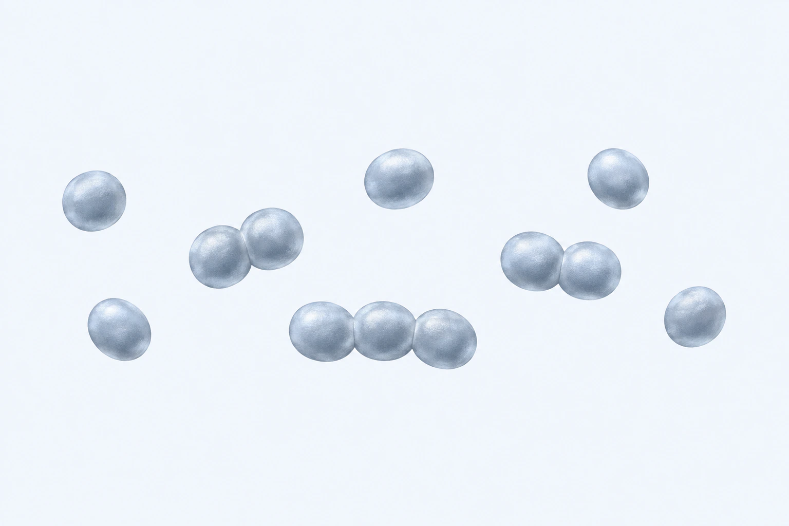
Type-strain-anchored Parvimonas micra smooth-morphotype reconstruction showing twelve coccoid bodies in five single, two paired, and one short-chain grouping. This species plate is representative, non-diagnostic, does not generalize surface structure to other morphotypes, and is not a micrograph.
Scientific media record1 verified identifier
- Subject
- Parvimonas micrataxon · species
- Identifiers
- NCBITaxon:33033
- Review
- Editorial review completeIdentifiers authority-verified · Accessibility validated · · parvimonas-micra|parvimonas-micra-morphology-v1.webp
- Digital source
- Trained-algorithmic mediaCreated with a trained generative algorithm and reviewed by WikiBiome for subject identity, scientific framing, identifiers, provenance, and accessibility.
- Scientific basis
- Parvimonas micra — NCBI TaxonomyParvimonas micra — LPSNProposal of Parvimonas gen. nov.Description of two morphotypes of Peptostreptococcus microsParvimonas micra type strain
- License
- CC BY-SA 4.0Created
Parvimonas micra is a small (0.5–1.0 µm), Gram-positive, obligately anaerobic coccus originally isolated from the human oral cavity that has emerged as one of the most consistent and universally enriched pathobionts in Colorectal Cancer across all geographic regions, age groups, and screening methodologies (16S rRNA gene, shotgun metagenomics, and qPCR).
Despite its status as a minor oral commensal, P. micra demonstrates remarkable CRC-specificity: its abundance is among the strongest single-taxon biomarkers for CRC across multiple large cohorts, suggesting it plays a direct or integrative role in colorectal carcinogenesis.
Its pathogenic mechanisms involve biofilm nucleation, iron piracy through siderophore production, adhesin-mediated epithelial contact, and integration into the polymicrobial oral-colorectal cancer consortium alongside Fusobacterium nucleatum, Parvimonas micra, and Clostridium symbiosum.
Contents
1. Taxonomy and Basic Properties2. Oral Origin and Translocation to the Colon3. Iron Dependency and Adhesin-Mediated Pathogenesis4. Biofilm Formation and Polymicrobial Consortia5. Pathogenic Mechanisms in Colorectal Cancer6. Disease Specificity and Biomarker Strength7. Detection and Quantification8. Typical Abundance Ranges9. Connections to WikiBiome Entities and Disease SignaturesTaxonomy and Basic Properties#
- Phylum: Firmicutes
- Class: Clostridia
- Order: Clostridiales
- Family: Peptoniphilaceae
- Genus: Parvimonas
- Species: Parvimonas micra (formerly Micromonas micra, Peptostreptococcus micra)
- Cell Type: Coccus (round); obligate anaerobe
- Cell Size: 0.5–1.0 µm diameter (among the smallest human microbiota bacteria; hence "parvi" = small)
- Gram Stain: Positive (thick peptidoglycan; no outer membrane)
- Motility: Non-motile; lacks flagella
- Genome: ~1.8 Mb (complete genome available)
- Spores: No spore formation; persists as vegetative cells
Oral Origin and Translocation to the Colon#
Oral Commensal-to-Pathobiont Transition#
P. micra is a normal inhabitant of the oral cavity, found in dental plaque, periodontal pockets, and the tongue dorsum in most healthy humans. In oral health, it occupies a minor ecological niche, antagonized by more dominant oral species (e.g., Streptococcus mutans, Prevotella intermedia).
However, under conditions of periodontal disease, poor oral hygiene, or Dysbiosis. P. micra increases in abundance within oral biofilms (10-fold expansion in periodontitis). Produces proteases and lipopolysaccharides (LPS) that damage periodontal tissues.
Translocates to the colon via a two-stage pathway.
Intestinal dysbiosis (reduced Butyrate, increased Metal-Driven Inflammation) compromises barrier integrity → increased gut permeability. Bloodstream translocation through the leaky epithelium; then reseeding into the colon via fecal circulation or direct recolonization.
Epidemiologically, patients with severe periodontitis have 2–3× higher CRC risk, supporting the oral-colorectal carcinoma axis hypothesis.
Iron Dependency and Adhesin-Mediated Pathogenesis#
Iron Acquisition via Siderophores#
P. micra is iron-dependent and produces catecholate-type siderophores to scavenge iron in the low-iron colonic environment.
Siderophore synthesis: Encodes genes for dihydroxybenzoic acid (DHBA) biosynthesis and iron-catecholate receptors. Mechanism: Secreted siderophores form tight iron(III) (Fe3+) complexes; the iron-siderophore complex is transported back via TonB-dependent receptors (similar to enterobactin piracy in E. coli).
Competitive effect: P. micra siderophores compete with host hepcidin-controlled iron, driving a local iron-scavenging microenvironment that advantages other iron-dependent pathogens (Fusobacterium, Bacteroides fragilis) while suppressing iron-sensitive commensals.
Adhesins and Epithelial Attachment#
P. micra produces multiple surface adhesins enabling direct epithelial contact:
| Adhesin | Target | Function |
|---|---|---|
| Type IV pili | Epithelial cell receptors (integrin α2β1, others) | Primary attachment; enable biofilm nucleation |
| Lipoteichoic acid (LTA) | TLR2 on epithelial and immune cells | PAMPs (pathogen-associated molecular patterns); trigger innate immune response |
| Outer surface proteins | Fibronectin, collagen, laminin | Extracellular matrix adhesion; especially in damaged epithelium |
These adhesins are particularly effective at sites of epithelial disruption—adenomatous polyps, ulcerated lesions, or dysbiotic low-butyrate areas where tight junctions are compromised.
Biofilm Formation and Polymicrobial Consortia#
P. micra is a skilled biofilm builder:
Biofilm Architecture#
- Produces polysaccharide matrix (exopolysaccharides; EPSs) that entraps other bacteria, creating a multi-species biofilm scaffold.
- Small cell size (0.5–1 µm) enables dense packing within biofilms; acts as a "nucleating core" for larger bacteria like Fusobacterium nucleatum and Bacteroides fragilis.
- Biofilm protects P. micra and partners from:
- Oxygen penetration (enabling strict anaerobes in micro-aerophilic colonic zones)
- Antibiotic and antimicrobial peptide penetration
- Neutrophil and immune cell attack
Oral-Colorectal Consortium Integration#
In CRC biofilms, P. micra integrates with:
| Partner | Role | Synergy |
|---|---|---|
| Fusobacterium nucleatum | FadA adhesin; invasin | F. nucleatum aggregates to P. micra biofilm core; together they breach epithelium |
| Parvimonas micra (above; listed for clarity) | Biofilm core; iron piracy | Nucleates polymicrobial biofilm; scavenges iron for all partners |
| Clostridium symbiosum | Bile acid metabolism; butyrate production | DCA/LCA-driven inflammation; muted butyrate in biofilm lowers pH |
| Peptostreptococcus stomatis | Colibactin (pks+ operon); genotoxin | DNA damage synergizes with P. micra adhesin-driven inflammation |
| Toxigenic Bacteroides fragilis (BFT+) | BFT toxin; barrier disruption | Synergistic epithelial damage; enable deeper biofilm invasion |
Pathogenic Mechanisms in Colorectal Cancer#
Direct Epithelial Invasion and Inflammation#
P. micra adheres directly to colonocyte apical surface via Type IV pili. Lipoteichoic acid (LTA) → TLR2 signaling → NF-κB activation → IL-6, IL-8, IL-1β production. Localized Th17 polarization (IL-17-producing CD4+ T cells) → further inflammation → epithelial damage.
Repeated cycles of adhesion, damage, and immune activation → chronic epithelial barrier compromise.
Integration with Colibactin and other Genotoxins#
Does not produce colibactin itself, but biofilm-integrated Peptostreptococcus stomatis (pks+) and Escherichia coli (pks+) strains do. P. micra biofilm acts as a delivery system for colibactin-producing partners, enabling direct epithelial contact with genotoxins. Colibactin → double-strand breaks in colonocytes → p53 activation, aberrant crypt formation, dysplasia.
Iron Dysbiosis and Functional Anemia#
P. micra iron siderophore production → local iron scarcity. Host responds with hepcidin elevation (systemic iron-withholding defense). Iron-dependent Faecalibacterium prausnitzii and butyrate producers → suppressed.
Iron-dependent pathogens (P. micra, Fusobacterium, Bacteroides fragilis) → enriched. Result: Functional iron anemia (low hepcidin paradoxically present with high circulating iron in heme/enterocyte-bound forms) drives CRC risk.
Disease Specificity and Biomarker Strength#
CRC-Specific Enrichment#
P. micra is one of the few taxa with CRC-specificity comparable to advanced cancer biomarkers.
Sensitivity: Detected in 70–95% of CRC cases (depending on polymerase chain reaction vs culture method). Specificity: Rare in healthy controls (<5% abundance); minimal in adenoma-only patients. Independence from screening method: Enriched in both 16S rRNA studies and metagenomics; independent biomarker strength despite methodological differences.
Cross-population consistency: Found in CRC cohorts across North America, Europe, Asia, and Africa.
Clinical Relevance#
Early CRC biomarker: Already enriched in advanced adenomas (AJCC stage III-IV); could enable early detection. Stage-independent: Abundance does not strongly correlate with TNM stage, suggesting P. micra enables adenoma-to-carcinoma transition rather than promoting late-stage progression.
Prognostic value: Emerging evidence suggests P. micra burden may correlate with poor prognosis and reduced response to immunotherapy.
Detection and Quantification#
Molecular Methods#
- 16S rRNA gene sequencing: Parvimonas micra-specific primers available; distinct from other Parvimonas spp.
- Shotgun metagenomics: P. micra genome is well-characterized; read abundance highly correlates with qPCR.
- qPCR: Species-specific assays; typical range:
- Healthy controls: <10^4 copies/g feces
- Adenoma patients: 10^5–10^7 copies/g feces
- CRC patients: 10^7–10^9 copies/g feces
Culture-Based Methods#
Anaerobic culture: Grows on Brucella agar + blood under 85% N2 / 10% H2 / 5% CO2. Colony morphology: Tiny (0.5–1 mm), translucent, mucoid colonies; slower growth than Fusobacterium. 16S rRNA RFLP or sequencing: Confirms identity; distinguishes from closely related Parvimonas species.
Biofilm Detection#
FISH (fluorescence in situ hybridization): Directly visualize P. micra within colorectal mucosal biofilms using species-specific probes. Confocal microscopy: Reveals P. micra biofilm structure and integration with Fusobacterium and other partners.
Typical Abundance Ranges#
| Population | P. micra (copies/g feces; % microbiota) | Notes |
|---|---|---|
| Healthy adults | <10^4 (<0.01%) | Minimal; oral carriage only |
| Periodontal disease patients | 10^5–10^6 (0.1–1%) | Elevated in mouth; may translocate |
| Adenoma patients (advanced) | 10^6–10^7 (0.5–2%) | Begin to enrich; biofilm formation |
| Incident CRC patients | 10^7–10^9 (2–10%) | Dramatically enriched; core biofilm member |
| Advanced CRC (stage III+) | 10^8–10^10 (5–15%) | Peak enrichment; strong biomarker |
Connections to WikiBiome Entities and Disease Signatures#
Iron – Absolute requirement; produces siderophores for iron piracy. – Catecholate-type iron-chelating compounds; compete with hepcidin. Hepcidin – Host iron-withholding defense; elevated in response to P. micra siderophore signaling.
– Type IV pili, LTA; direct epithelial attachment. – PAMP; TLR4 signaling. – PAMP; TLR2 signaling (Gram-positive).
Biofilm – Major biofilm nucleator; core structural component of polymicrobial CRC biofilms. Colorectal Cancer – One of the strongest single-taxon biomarkers; present in 70–95% of CRC cases. – Originates in oral cavity; translocates to colon; member of oral pathobiont consortium.
– Enriched in periodontal disease; periodontal disease patients have 2–3× higher CRC risk. Fusobacterium nucleatum – cobalt (Co)-enriched with P. micra; biofilm integration. Peptostreptococcus stomatis – cobalt-enriched; synergistic genotoxin delivery.
Clostridium symbiosum – cobalt-enriched; synergistic inflammation. Bacteroides fragilis (especially BFT+ strains) – cobalt-enriched; synergistic barrier disruption. inflammation – TLR2-driven NF-κB activation; Th17 polarization.
dysbiosis – Enriched in dysbiotic CRC microbiota; suppressed in healthy, butyrate-dominated microbiota. Faecalibacterium prausnitzii – Inverse relationship; suppressed where P. micra iron-scavenging dominates.
Parvimonas micra exemplifies the oral-colorectal axis: a minor oral commensal that becomes a major CRC biomarker upon translocation to the dysbiotic colon, where its biofilm-nucleating and iron-scavenging capabilities integrate it into a polymicrobial carcinogenic consortium.
References 6
Numbered by first appearance in the article, then reconciled with its declared source list.
- 1
Youwen Qin, Xin Tong, Wei-Jian Mei et al. (2024). Consistent signatures in the human gut microbiome of old- and young-onset colorectal cancer. Nature Communications.
- 2
Bars-Cortina D, Ramon E, Rius-Sansalvador B et al. (2024). Comparison between 16S rRNA and Shotgun Sequencing in Colorectal Cancer, Advanced Colorectal Lesions, and Healthy Human Gut Microbiota. BMC Genomics.
- 3
Wu Y, Jiao N, Zhu R et al. (2021). Identification of Microbial Markers across Populations in Early Detection of Colorectal Cancer. Nature Communications.
- 4
Lan Zhao, William C. Cho, Mark R. Nicolls (2021). Colorectal cancer-associated microbiome patterns and signatures. Frontiers in Genetics.
- 5
Hou X, Zheng Z, Wei J et al. (2022). Effects of Gut Microbiota on Immune Responses and Immunotherapy in Colorectal Cancer. Frontiers in Immunology.
- 6
Osman MA, Neoh HM, Ab Mutalib NS et al. (2018). 16S rRNA Gene Sequencing for Deciphering the Colorectal Cancer Gut Microbiome: Current Protocols and Workflows. Frontiers in Microbiology.
Article network
Mentioned here 15
Pages linking here 3
Connect the evidence
Publicly readable discussion by ORCID-authenticated researchers. Questions, interpretation, methods, corrections, and new evidence stay attached to this record.
No discussion yet. Start with a precise question or a source-backed observation.
Activity and accepted changes
Accepted researcher context, editorial status, public discussion, and upstream Git revisions are shown together. Pending, declined, and withdrawn proposals remain private.
- published revision
Backfill inflammation concept links
Karen Pendergrass · +2 −2
Inspect exact Git diff ↗ - published revision
Complete corpus-wide Dysbiosis linking
Karen Pendergrass · +2 −2
Inspect exact Git diff ↗ - published revision
Complete Tight junctions contextual coverage
Karen Pendergrass · +1 −1
Inspect exact Git diff ↗ - published revision
massive wiki expansion: 149 stubs fixed, 100+ new pages, Rule 15 scan, keystone papers
WikiBiome Deploy Bot · +78 −74
Inspect exact Git diff ↗ - published revision
nightly maintenance: 94 stub demotions, 181 source_count fixes, 22 auto-discovered stubs, 5 adversarial audits, 3 boundary fixes, 3 evidence-level corrections
WikiBiome Deploy Bot · +3 −0
Inspect exact Git diff ↗ - published revision
WikiBiome update — 2026-04-15 17:23
WikiBiome Deploy Bot · +15 −15
Inspect exact Git diff ↗ - published revision
wiki: bulk entity upgrades, new article pages, and site regeneration
WikiBiome Deploy Bot · +6 −0
Inspect exact Git diff ↗ - published revision
WikiBiome update — integrity fixes, metallomic diet pages, cross-condition analyses
WikiBiome Deploy Bot · +30 −28
Inspect exact Git diff ↗ - published revision
WikiBiome update — 2026-04-10 17:16
WikiBiome Deploy Bot · +185 −0
Inspect exact Git diff ↗