
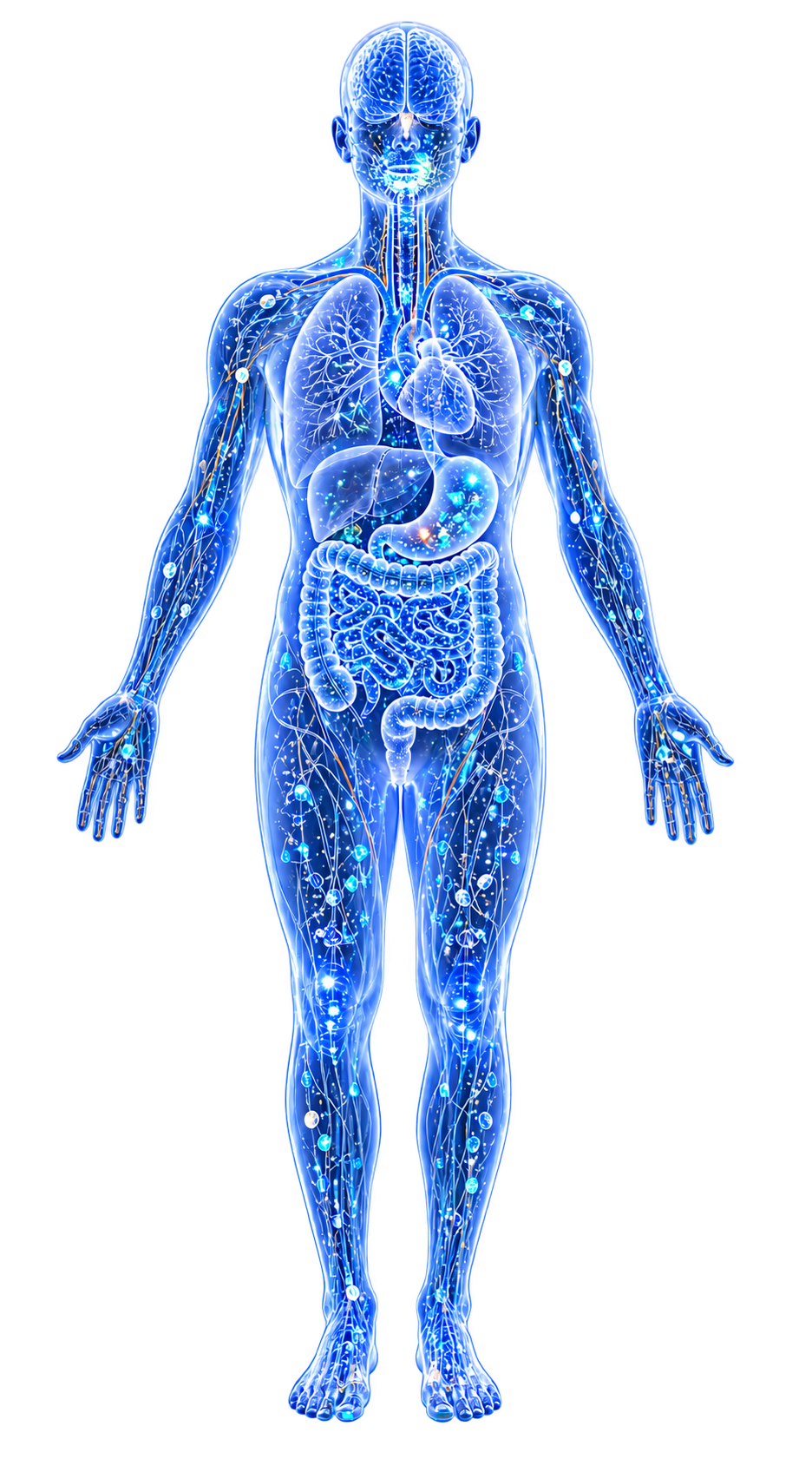
This page unifies the infant formula contamination, pregnancy complications, autism spectrum disorder, necrotizing enterocolitis, and neurodevelopmental toxicity threads around a single organizing insight: metal toxicity is disproportionately severe during specific developmental windows, and much of the damage sustained during these windows is irreversible.
The developing organism is not simply a small adult. It differs qualitatively in barrier integrity, detoxification capacity, absorption kinetics, and the vulnerability of the biological processes underway at each stage. Recognizing these differences demands developmental-stage-specific risk assessment—something the current regulatory framework largely fails to provide.
Evidence map65 cited passagesInspect provenance +
Immature detoxification systems: Low metallothionein expression, undeveloped hepatic phase II conjugation pathways, and lower baseline glutathione levels mean that infants and young children cannot neutralize or excrete metals as efficiently as adults, ].
Higher intake per body weight: A 6-month-old infant consuming 800 mL of formula weighs approximately 7-8 kg. The same absolute quantity of a contaminant that would represent a negligible dose for a 70 kg adult becomes a physiologically significant exposure when normalized to body mass. Commercial vegetable purees further concentrate contaminants from substan
Incomplete biological barriers: The blood-brain barrier (BBB) is not fully formed until approximately age 2-3, allowing metals like lead, mercury, and aluminum direct access to the developing brain, ]. The intestinal barrier is more permeable in neonates, particularly preterm infants. The placental barrier is compromised by common pregnancy complications inc
Rapid cell division: The high mitotic rate in developing tissues creates more targets for genotoxic and epigenetic damage. Metals that interfere with DNA methylation, histone modification, or chromosomal integrity during periods of rapid proliferation can produce effects that propagate through all daughter cells.
Active neurogenesis and synaptogenesis: The prenatal-through-early-childhood period encompasses the most intensive phases of neural development—neuronal migration, axon pathfinding, synapse formation, myelination, and pruning. These processes depend on zinc-dependent scaffolding proteins (SHANK3, NLGN-NRXN complexes), calcium-dependent signaling, and iron
Irreversibility of developmental damage: Unlike adult-onset toxicity, where cessation of exposure may allow recovery, damage sustained during critical developmental windows often produces permanent structural and functional deficits. A neuron that fails to migrate to its correct position, a synapse that fails to form, or an epigenetic mark established incorr
Additionally, the higher gastrointestinal absorption rates in infants amplify effective exposure: lead absorption is estimated at 30-50% in children versus 5-15% in adults—a 3-10 fold difference in bioavailability from the same dietary concentration, ].
Placental barrier compromise in pregnancy complications: The placenta normally restricts nickel transfer to the fetus, but this barrier function is degraded by common pregnancy complications. In a Chinese case-control study (n=72), Ding et al. (2021) found that cord blood nickel was significantly elevated in women with gestational diabetes mellitus (GDM) and
Congenital heart defects: Zhang et al. (2019) demonstrated dose-dependent associations between maternal nickel exposure and congenital heart defect (CHD) risk in a Chinese case-control study (n=889). Hair nickel was significantly elevated in CHD cases (0.629 vs. 0.443 ng/mg, P<.001), and placental nickel was also elevated (0.178 vs. 0.148 ng/mg, P=.039). The
Prenatal lead exposure and child gut microbiome: Eggers et al. (2023) found that prenatal lead exposure during pregnancy was negatively associated with the gut microbiome composition in children aged 9-11 years—effects persisting nearly a decade after the exposure window. Second trimester Pb exposure showed the strongest negative association, supporting t
Prenatal metal mixtures and maternal mental health: In the Project Viva cohort (n=1,226), Rokoff et al. (2023) found weak but consistent associations between first-trimester erythrocyte lead and elevated depressive symptoms during pregnancy (OR 1.19 per doubling of Pb). While the overall metal mixture effect was null in this relatively well-nourished cohort,
Epigenetic programming: The prenatal period is when the developmental epigenome is being actively established. Metals that disrupt DNA methylation and histone modification during this window can create permanent epigenetic changes that alter gene expression across the lifespan. Lead promotes amyloid-beta accumulation through APP gene demethylation—an effe
Zinc deficiency during pregnancy: Zinc deficiency during pregnancy causes ASD-like behavior in animal models, while prenatal zinc supplementation prevents VPA-induced ASD-like behaviors. Approximately 10% of the human genome encodes zinc-binding proteins; toxic metals (Pb, Hg, Cd) compete with zinc for these binding sites, effectively creating functional zin
United Kingdom: All 24 prescription infant formulas tested were contaminated with aluminum, ranging from 41-1956 ug/L. The worst offender (Abbott PediaSure Plus Juice Apple) contained 1956 ug/L—391 ug per serving. Breast milk, by contrast, has aluminum content an order of magnitude lower.
Germany: The most comprehensive European total diet study found infant formula contributed up to 64% of total dietary exposure for some contaminants in infants under 1 year. Cadmium exceeded health-based guidance values for approximately 30% of infants. Nickel exposure was 2.96-3.54 ug/kg bw/day. The margin of exposure for inorganic arsenic was below 1 for a
United States: All 10 commercial baby food products from Houston tested positive for heavy metals. Aluminum (up to 4.09 ug/g) and zinc exceeded ATSDR minimal risk levels. Arsenic was detected in all infant formulas tested, with arsenic in formula almost exclusively in the inorganic (more toxic) form, ].
Spain: Baby food jars showed nickel reaching 79-86% of TDI at standard consumption (130 g/day for a 9 kg infant). Aluminum intake from vegetable jars reached 55-160% of TWI. Manganese intake was 40 times higher than recommended values.
Italy: Among toxic elements in Italian baby foods, nickel showed the highest daily intake at 9.43 ug/kg bw/day in one powdered milk sample, representing approximately 85.7% of the PTWI-derived daily risk estimator.
Soy-based formulas carry elevated risk: Soy-based infant formulas have higher nickel concentrations than milk-based formulas (0.45 vs. 0.03 mg/L Ni—approximately 10-fold higher), and orders of magnitude more than human breast milk (0.005-0.016 mg/L), ].
The concentration effect: Commercial vegetable purees deliver the nutritional and contaminant content of substantially larger quantities of fresh vegetables, concentrated into a serving consumed by a body 10-15 times smaller than an adult. The industry shift from small flavor-learning portions to concentrated vegetable fortification occurred without toxicoki
Nickel and necrotizing enterocolitis (NEC): Pendergrass (2026) proposes that dietary nickel from infant formula may be a critical but overlooked contributor to NEC pathogenesis in preterm infants. The key NEC-associated pathogens—E. coli, Klebsiella, Enterobacter, Citrobacter, Ureaplasma—all rely on nickel-dependent enzymes: urease, [NiFe]-hydrogenase,
EFSA TDI exceedance: The EFSA tolerable daily intake for nickel (13 ug/kg BW) may be exceeded by a substantial fraction of formula-fed infants. The German total diet study found nickel exposure of 2.96-3.54 ug/kg bw/day at median intake, with high-consuming toddlers reaching 17.9 ug/kg bw/day—exceeding the TDI.
Dietary TDI exceedances are common: A French study of children aged 1-36 months found that 7.9-37.9% exceeded the nickel TDI under lower-bound assumptions, and under upper-bound assumptions, up to 98% exceeded TDI. Chocolate and cocoa products accounted for 10% of mean daily nickel intake in French children.
Main dietary sources overlap with children's preferences: The foods richest in nickel—chocolate, cocoa, cereals, nuts, and legumes—are consumed heavily by young children. Cocoa contains 8.2-17.1 mg Ni/kg; grains, legumes, and cereals contain 0.3-9.8 mg Ni/kg.
Showing 24 of 65 evidence-bearing passages. Every remaining citation is still indexed in the reference record below.
Contents
1. 1. The Principle2. 2. Critical Windows3. 3. Mechanisms of Enhanced Vulnerability4. 4. The Regulatory Gap5. 5. Connections1. The Principle#
Developing organisms—from embryos through early childhood—are disproportionately vulnerable to metal toxicity for six converging reasons:
- Immature detoxification systems: Low metallothionein expression, undeveloped hepatic phase II conjugation pathways, and lower baseline glutathione levels mean that infants and young children cannot neutralize or excrete metals as efficiently as adults.[1]Nickel Exposure from Food and Levels in Children's Blood and Tissues: Health ImplicationsDobrzyńska MM, Gajowik A, Jankowska-Steifer EA et al. · 2025Open reference 1 ↓[2]Toxicity, Mechanism and Health Effects of Some Heavy MetalsMonisha Jaishankar, Tenzin Tseten, Naresh Anbalagan et al. · 2014Open reference 2 ↓
- Higher intake per body weight: A 6-month-old infant consuming 800 mL of formula weighs approximately 7-8 kg. The same absolute quantity of a contaminant that would represent a negligible dose for a 70 kg adult becomes a physiologically significant exposure when normalized to body mass. Commercial vegetable purees further concentrate contaminants from substantially larger quantities of fresh vegetables into servings consumed by a body 10-15 times smaller than an adult.[3]Age-Window Metabolic and Toxicokinetic Vulnerability in Vegetable-Based Baby Foods: Separating Developmental Readiness from Toxicant SusceptibilityKaren Pendergrass · 2026Open reference 3 ↓
- Incomplete biological barriers: The blood-brain barrier (BBB) is not fully formed until approximately age 2-3, allowing metals like lead, mercury, and aluminum direct access to the developing brain.[4]Interaction of Heavy Metal Lead with Gut Microbiota: Implications for Autism Spectrum DisorderTizabi Y, Bennani S, El Kouhen N et al. · 2023Open reference 4 ↓[5]Metal Profiles in Autism Spectrum Disorders: A Crosstalk between Toxic and Essential MetalsBlazewicz A, Grabrucker AM · 2023Open reference 5 ↓ The intestinal barrier is more permeable in neonates, particularly preterm infants. The placental barrier is compromised by common pregnancy complications including gestational diabetes and hypertensive disorders.[6]Pregnancy complications effect on the nickel content in maternal blood, placenta blood and umbilical cord blood during pregnancyDing R, Ruan Y, He X et al. · 2021Open reference 6 ↓
- Rapid cell division: The high mitotic rate in developing tissues creates more targets for genotoxic and epigenetic damage. Metals that interfere with DNA methylation, histone modification, or chromosomal integrity during periods of rapid proliferation can produce effects that propagate through all daughter cells.[7]Metal nickel exposure increase the risk of congenital heart defects occurrence in offspringZhang N, Chen W, Dong Y et al. · 2019Open reference 7 ↓
- Active neurogenesis and synaptogenesis: The prenatal-through-early-childhood period encompasses the most intensive phases of neural development—neuronal migration, axon pathfinding, synapse formation, myelination, and pruning. These processes depend on zinc-dependent scaffolding proteins (SHANK3, NLGN-NRXN complexes), calcium-dependent signaling, and iron-dependent myelination—all of which are disrupted by toxic metals.[5]Metal Profiles in Autism Spectrum Disorders: A Crosstalk between Toxic and Essential MetalsBlazewicz A, Grabrucker AM · 2023Open reference 5 ↓[4]Interaction of Heavy Metal Lead with Gut Microbiota: Implications for Autism Spectrum DisorderTizabi Y, Bennani S, El Kouhen N et al. · 2023Open reference 4 ↓
- Irreversibility of developmental damage: Unlike adult-onset toxicity, where cessation of exposure may allow recovery, damage sustained during critical developmental windows often produces permanent structural and functional deficits. A neuron that fails to migrate to its correct position, a synapse that fails to form, or an epigenetic mark established incorrectly during organogenesis cannot be corrected later.[3]Age-Window Metabolic and Toxicokinetic Vulnerability in Vegetable-Based Baby Foods: Separating Developmental Readiness from Toxicant SusceptibilityKaren Pendergrass · 2026Open reference 3 ↓
Additionally, the higher gastrointestinal absorption rates in infants amplify effective exposure: lead absorption is estimated at 30-50% in children versus 5-15% in adults—a 3-10 fold difference in bioavailability from the same dietary concentration.[3]Age-Window Metabolic and Toxicokinetic Vulnerability in Vegetable-Based Baby Foods: Separating Developmental Readiness from Toxicant SusceptibilityKaren Pendergrass · 2026Open reference 3 ↓[2]Toxicity, Mechanism and Health Effects of Some Heavy MetalsMonisha Jaishankar, Tenzin Tseten, Naresh Anbalagan et al. · 2014Open reference 2 ↓
2. Critical Windows#
Prenatal / Placental#
The prenatal period represents the first and arguably most consequential vulnerability window. The developing embryo and fetus depend entirely on placental transfer for nutrition and are exposed to whatever metals cross the placental barrier.
Placental barrier compromise in pregnancy complications: The placenta normally restricts nickel transfer to the fetus, but this barrier function is degraded by common pregnancy complications.
In a Chinese case-control study (n=72), Ding et al. (2021) found that cord blood nickel was significantly elevated in women with gestational diabetes mellitus (GDM) and hypertensive disorders complicating pregnancy (HDCP) versus controls. The barrier function ranking from best to worst: Control > GDM > Disease combination (GDM+HDCP) > HDCP.
In controls, 85% maintained a maternal:cord blood nickel (Ni) ratio >1 (indicating effective barrier function), compared to only 50-60% in disease groups. Birth weight and length were significantly reduced in the HDCP group.[6]Pregnancy complications effect on the nickel content in maternal blood, placenta blood and umbilical cord blood during pregnancyDing R, Ruan Y, He X et al. · 2021Open reference 6 ↓
Congenital heart defects: Zhang et al. (2019) demonstrated dose-dependent associations between maternal nickel exposure and congenital heart defect (CHD) risk in a Chinese case-control study (n=889). Hair nickel was significantly elevated in CHD cases (0.629 vs. 0.443 ng/mg, P<.001), and placental nickel was also elevated (0.178 vs.
0.148 ng/mg, P=.039). The highest tertile of hair nickel produced adjusted odds ratios of 1.326 for total CHDs, with stronger associations for specific subtypes: left ventricular outflow tract obstruction (aOR 1.549), right ventricular outflow tract obstruction (aOR 1.543), and septal defects (aOR 1.443).[7]Metal nickel exposure increase the risk of congenital heart defects occurrence in offspringZhang N, Chen W, Dong Y et al. · 2019Open reference 7 ↓
Prenatal lead exposure and child Gut Microbiome: Eggers et al. (2023) found that prenatal lead exposure during pregnancy was negatively associated with the gut microbiome composition in children aged 9-11 years—effects persisting nearly a decade after the exposure window.
Second trimester lead (Pb) exposure showed the strongest negative association, supporting the second trimester as a critical window. Key taxa negatively associated with prenatal lead included Bacteroides caccae, Bifidobacterium longum, and Bifidobacterium bifidum—organisms important for immune development and gut health.[8]Prenatal Lead Exposure is Negatively Associated with the Gut Microbiome in ChildhoodEggers S, Midya V, Bixby M et al. · 2023Open reference 8 ↓
Prenatal metal mixtures and maternal mental health: In the Project Viva cohort (n=1,226), Rokoff et al. (2023) found weak but consistent associations between first-trimester erythrocyte lead and elevated depressive symptoms during pregnancy (OR 1.19 per doubling of lead).
While the overall metal mixture effect was null in this relatively well-nourished cohort, the findings suggest that higher-exposure populations may face compounded mental health risks during pregnancy.[9]Early pregnancy essential and non-essential metal mixtures and maternal antepartum and postpartum depressive symptomsRokoff LB, Cardenas A, Lin PI et al. · 2023Open reference 9 ↓
Epigenetic programming: The prenatal period is when the developmental epigenome is being actively established. Metals that disrupt DNA methylation and histone modification during this window can create permanent epigenetic changes that alter gene expression across the lifespan.
Lead promotes amyloid-beta accumulation through APP gene demethylation—an effect of early-life exposure that manifests as Alzheimer's-related pathology decades later.[5]Metal Profiles in Autism Spectrum Disorders: A Crosstalk between Toxic and Essential MetalsBlazewicz A, Grabrucker AM · 2023Open reference 5 ↓
Nickel induces epigenetic alterations including changes in DNA methylation and histone acetylation, proposed as a mechanism for cardiac teratogenesis.[7]Metal nickel exposure increase the risk of congenital heart defects occurrence in offspringZhang N, Chen W, Dong Y et al. · 2019Open reference 7 ↓
Zinc deficiency during pregnancy: Zinc deficiency during pregnancy causes ASD-like behavior in animal models, while prenatal zinc supplementation prevents VPA-induced ASD-like behaviors.
Approximately 10% of the human genome encodes zinc-binding proteins; toxic metals (lead, mercury (Hg), cadmium (Cd)) compete with zinc for these binding sites, effectively creating functional zinc deficiency during the most critical period of brain development.[5]Metal Profiles in Autism Spectrum Disorders: A Crosstalk between Toxic and Essential MetalsBlazewicz A, Grabrucker AM · 2023Open reference 5 ↓
> See also: Nickel and Reproductive/Developmental Toxicity for detailed coverage of placental nickel toxicology.
Neonatal / Infant Formula#
The neonatal period introduces the second major vulnerability window. Infants who are not exclusively breastfed are exposed to whatever contaminants are present in their formula—their sole or primary nutritional source during the period of most rapid postnatal brain growth.
Infant formula contamination is widespread and global: Studies across at least seven countries document consistent multi-element contamination of infant formulas and baby foods.
United Kingdom: All 24 prescription infant formulas tested were contaminated with aluminum, ranging from 41-1956 ug/L. The worst offender (Abbott PediaSure Plus Juice Apple) contained 1956 ug/L—391 ug per serving. Breast milk, by contrast, has aluminum content an order of magnitude lower.[10]Prescription Infant Formulas Are Contaminated with AluminiumRedgrove J, Rodriguez I, Mahadevan-Bava S et al. · 2019Open reference 10 ↓
Germany: The most comprehensive European total diet study found infant formula contributed up to 64% of total dietary exposure for some contaminants in infants under 1 year. Cadmium exceeded health-based guidance values for approximately 30% of infants. Nickel exposure was 2.96-3.54 ug/kg bw/day.
The margin of exposure for inorganic arsenic was below 1 for all children, indicating potential cancer risk.[11]The contribution of infant formula to the food survey-based dietary exposure of nine selected elementsHopfner T, Wollenberg M, Jager A et al. · 2025Open reference 11 ↓
United States: All 10 commercial baby food products from Houston tested positive for Heavy Metals. Aluminum (up to 4.09 ug/g) and zinc exceeded ATSDR minimal risk levels.
Arsenic was detected in all infant formulas tested, with arsenic in formula almost exclusively in the inorganic (more toxic) form.[12]Evaluation of Heavy Metals in Commercial Baby FoodsGaruba OD, Anglin JC, Good S et al. · 2024Open reference 12 ↓[13]Arsenic concentration and speciation in infant formulas and first foodsJackson BP, Taylor VF, Punshon T et al. · 2012Open reference 13 ↓
Spain: Baby food jars showed nickel reaching 79-86% of TDI at standard consumption (130 g/day for a 9 kg infant). Aluminum intake from vegetable jars reached 55-160% of TWI. Manganese intake was 40 times higher than recommended values.[14]Baby Food Jars as a Dietary Source of Essential (K, Na, Ca, Mg, Fe, Zn, Cu, Co, Mo, Mn) and Toxic Elements (Al, Cd, Pb, B, Ba, V, Sr, Li, Ni)Gonzalez-Suarez S, Paz-Montelongo S, Niebla-Canelo D et al. · 2022Open reference 14 ↓
Italy: Among toxic elements in Italian baby foods, nickel showed the highest daily intake at 9.43 ug/kg bw/day in one powdered milk sample, representing approximately 85.7% of the PTWI-derived daily risk estimator.[15]Chemical characterization of baby food consumed in ItalyMeli MA, Desideri D, Sisti D et al. · 2024Open reference 15 ↓
Soy-based formulas carry elevated risk: Soy-based infant formulas have higher nickel concentrations than milk-based formulas (0.45 vs. 0.03 mg/L nickel (Ni)—approximately 10-fold higher), and orders of magnitude more than human breast milk (0.005-0.016 mg/L).[16]Nickel as a Catalytic Driver of Necrotizing Enterocolitis: Dietary Nickel, Microbial Metallomics, and the Activation of Nickel-Dependent Virulence Pathways in the Preterm GutKaren Pendergrass · 2026Open reference 16 ↓[1]Nickel Exposure from Food and Levels in Children's Blood and Tissues: Health ImplicationsDobrzyńska MM, Gajowik A, Jankowska-Steifer EA et al. · 2025Open reference 1 ↓
The concentration effect: Commercial vegetable purees deliver the nutritional and contaminant content of substantially larger quantities of fresh vegetables, concentrated into a serving consumed by a body 10-15 times smaller than an adult.
The industry shift from small flavor-learning portions to concentrated vegetable fortification occurred without toxicokinetic evaluation.[3]Age-Window Metabolic and Toxicokinetic Vulnerability in Vegetable-Based Baby Foods: Separating Developmental Readiness from Toxicant SusceptibilityKaren Pendergrass · 2026Open reference 3 ↓
Nickel and necrotizing enterocolitis (NEC): Pendergrass (2026) proposes that dietary nickel from infant formula may be a critical but overlooked contributor to NEC pathogenesis in preterm infants. The key NEC-associated pathogens—E. coli, Klebsiella, Enterobacter, Citrobacter, Ureaplasma—all rely on nickel-dependent enzymes: Urease, [NiFe]-Hydrogenase, and Glyoxalase I I.
Excess dietary nickel creates a positive feedback loop: nickel-fueled urease raises gut pH, favoring Proteobacteria over acid-producing commensals like Lactobacillus. Human breast milk is naturally nickel-poor, potentially as an evolved mechanism of Nutritional Immunity (Metal Sequestration)—starving nickel-dependent pathogens of their essential cofactor.
Formula feeding may overwhelm the host's calprotectin and lactoferrin metal-sequestration defenses, enabling pathogen virulence.[16]Nickel as a Catalytic Driver of Necrotizing Enterocolitis: Dietary Nickel, Microbial Metallomics, and the Activation of Nickel-Dependent Virulence Pathways in the Preterm GutKaren Pendergrass · 2026Open reference 16 ↓
EFSA TDI exceedance: The EFSA tolerable daily intake for nickel (13 ug/kg BW) may be exceeded by a substantial fraction of formula-fed infants.
The German total diet study found nickel exposure of 2.96-3.54 ug/kg bw/day at median intake, with high-consuming toddlers reaching 17.9 ug/kg bw/day—exceeding the TDI.[11]The contribution of infant formula to the food survey-based dietary exposure of nine selected elementsHopfner T, Wollenberg M, Jager A et al. · 2025Open reference 11 ↓
Early Childhood (1-5 years)#
The transition to solid foods introduces new exposure routes, while the vulnerability factors of immaturity persist.
Dietary TDI exceedances are common: A French study of children aged 1-36 months found that 7.9-37.9% exceeded the nickel TDI under lower-bound assumptions, and under upper-bound assumptions, up to 98% exceeded TDI.
Chocolate and cocoa products accounted for 10% of mean daily nickel intake in French children.[1]Nickel Exposure from Food and Levels in Children's Blood and Tissues: Health ImplicationsDobrzyńska MM, Gajowik A, Jankowska-Steifer EA et al. · 2025Open reference 1 ↓
Main dietary sources overlap with children's preferences: The foods richest in nickel—chocolate, cocoa, cereals, nuts, and legumes—are consumed heavily by young children. Cocoa contains 8.2-17.1 mg nickel (Ni)/kg; grains, legumes, and cereals contain 0.3-9.8 mg nickel/kg.[1]Nickel Exposure from Food and Levels in Children's Blood and Tissues: Health ImplicationsDobrzyńska MM, Gajowik A, Jankowska-Steifer EA et al. · 2025Open reference 1 ↓
Industrial proximity amplifies risk: Children living near industrial areas (e.g., petrochemical facilities) have elevated urinary nickel correlated with markers of Oxidative Stress, demonstrating that environmental exposure compounds dietary intake.[1]Nickel Exposure from Food and Levels in Children's Blood and Tissues: Health ImplicationsDobrzyńska MM, Gajowik A, Jankowska-Steifer EA et al. · 2025Open reference 1 ↓
Nickel sensitization begins early: Nickel allergy prevalence is already 8-10% in children, increasing with ear piercing and other metal contact. Early sensitization establishes a lifelong immunological vulnerability—nickel-sensitized individuals develop eczematous flare-ups at dietary intakes above the LOAEL of 4.3 ug nickel/kg body weight.[1]Nickel Exposure from Food and Levels in Children's Blood and Tissues: Health ImplicationsDobrzyńska MM, Gajowik A, Jankowska-Steifer EA et al. · 2025Open reference 1 ↓
Lead exposure during this window has irreversible neurodevelopmental effects: Blood lead levels above 10 ug/dL affect IQ in children. Lead crosses the developing BBB more readily than the mature barrier, mimics calcium in signaling pathways critical for synaptogenesis, and produces latent epigenetic changes that manifest as neurodegeneration decades later.
There is no established safe threshold for lead exposure in children.[2]Toxicity, Mechanism and Health Effects of Some Heavy MetalsMonisha Jaishankar, Tenzin Tseten, Naresh Anbalagan et al. · 2014Open reference 2 ↓[4]Interaction of Heavy Metal Lead with Gut Microbiota: Implications for Autism Spectrum DisorderTizabi Y, Bennani S, El Kouhen N et al. · 2023Open reference 4 ↓
Neurodevelopmental Window (Prenatal through ~Age 6)#
The neurodevelopmental vulnerability window spans the prenatal period through approximately age 6, encompassing neuronal proliferation, migration, differentiation, synaptogenesis, myelination, and synaptic pruning. This extended window is where the pregnancy, infant formula, and autism threads converge.
Autism spectrum disorder and metal dyshomeostasis: A growing body of evidence documents consistent metal signatures in ASD.
Across 25+ studies using hair, blood, urine, and teeth samples, the pattern is remarkably consistent: toxic metals (lead (Pb), mercury (Hg), cadmium (Cd)) are elevated while essential metals (especially zinc (Zn)) are depleted in ASD children.[5]Metal Profiles in Autism Spectrum Disorders: A Crosstalk between Toxic and Essential MetalsBlazewicz A, Grabrucker AM · 2023Open reference 5 ↓
Essential metal depletion: Iron and zinc are significantly lower in ASD children.
Zinc deficiency during brain development may be a primary pathological event, with toxic metals creating functional zinc deficiency by competing for the same binding sites—approximately 10% of the human genome encodes zinc-binding proteins.[5]Metal Profiles in Autism Spectrum Disorders: A Crosstalk between Toxic and Essential MetalsBlazewicz A, Grabrucker AM · 2023Open reference 5 ↓
Gut pathology as a mediating mechanism: O'Grady and Grabrucker (2025) systematically demonstrated that heavy metal exposure (mercury, cadmium, lead) and zinc deficiency produce overlapping gut pathologies: intestinal barrier dysfunction, increased permeability, gut Metal-Driven Inflammation, structural damage, and microbiota Dysbiosis.
Notably, 30-70% of children with ASD suffer from GI disturbance, far exceeding rates in the general pediatric population, and GI symptom severity correlates with ASD behavioral severity.[17]Metal Dyshomeostasis as a Driver of Gut Pathology in Autism Spectrum DisordersO'Grady K, Grabrucker AM · 2025Open reference 17 ↓
Toxic metals predict social behavior variance in typical children: Heavy metal load in urine accounted for 32% of variance in social behavior outcomes even in neurotypical children, demonstrating that metal effects on neurodevelopment are not binary (ASD vs. no ASD) but operate along a continuum of severity.[3]Age-Window Metabolic and Toxicokinetic Vulnerability in Vegetable-Based Baby Foods: Separating Developmental Readiness from Toxicant SusceptibilityKaren Pendergrass · 2026Open reference 3 ↓
Synaptogenesis vulnerability through SHANK3: The SHANK3 scaffold protein, critical for postsynaptic density organization, requires zinc for proper function. SHANK3 participates in the NLGN-NRXN-SHANK synaptic complex, which is modified by both zinc and calcium and is disrupted by toxic metals that compete for these binding sites.
Mutations in SHANK3 are among the most common monogenic causes of ASD, and zinc depletion via toxic metal competition may functionally mimic these genetic disruptions during the critical window of synaptogenesis.[5]Metal Profiles in Autism Spectrum Disorders: A Crosstalk between Toxic and Essential MetalsBlazewicz A, Grabrucker AM · 2023Open reference 5 ↓
Lead-gut-brain axis in ASD: Lead disrupts the gut-brain axis through multiple converging pathways: inducing gut dysbiosis (increased Firmicutes and Bacteroidetes, decreased Proteobacteria), damaging the intestinal barrier (reduced MUC2, ZO-1, claudin-1, occludin), crossing the immature BBB, displacing calcium from neurotransmitter release machinery, activating microglia and driving neuroinflammation, and damaging oligodendrocytes to impair myelination.
Even low blood lead concentrations at age 7-8 are associated with more autistic behaviors at age 11-12.[4]Interaction of Heavy Metal Lead with Gut Microbiota: Implications for Autism Spectrum DisorderTizabi Y, Bennani S, El Kouhen N et al. · 2023Open reference 4 ↓
Mercury neurotoxicity in the developmental window: Mercury, particularly methylmercury, readily crosses both the BBB and the placental barrier. An estimated 8-10% of American women have mercury levels that could induce neurological disorders in their children.
methylmercury downregulates myelin basic protein expression, contributing to demyelination during the critical myelination window of early childhood.[5]Metal Profiles in Autism Spectrum Disorders: A Crosstalk between Toxic and Essential MetalsBlazewicz A, Grabrucker AM · 2023Open reference 5 ↓[2]Toxicity, Mechanism and Health Effects of Some Heavy MetalsMonisha Jaishankar, Tenzin Tseten, Naresh Anbalagan et al. · 2014Open reference 2 ↓
3. Mechanisms of Enhanced Vulnerability#
Immature Barriers#
Three barrier systems that protect the adult organism from metal toxicity are incompletely formed or functionally compromised during development.
Blood-brain barrier: Not fully mature until approximately age 2-3. The developing BBB allows metals including lead, mercury, and aluminum direct access to the brain during the most intensive period of neurogenesis and synaptogenesis.[4]Interaction of Heavy Metal Lead with Gut Microbiota: Implications for Autism Spectrum DisorderTizabi Y, Bennani S, El Kouhen N et al. · 2023Open reference 4 ↓
Intestinal barrier: More permeable in neonates, particularly preterm infants. This increased permeability allows greater systemic absorption of ingested metals and permits bacterial translocation—the latter being directly relevant to NEC pathogenesis.[16]Nickel as a Catalytic Driver of Necrotizing Enterocolitis: Dietary Nickel, Microbial Metallomics, and the Activation of Nickel-Dependent Virulence Pathways in the Preterm GutKaren Pendergrass · 2026Open reference 16 ↓
Placental barrier: Normally restricts metal transfer to the fetus, but is compromised by GDM and HDCP. In hypertensive disorders, only 50-60% of women maintain effective placental barrier function for nickel, compared to 85% in healthy pregnancies.[6]Pregnancy complications effect on the nickel content in maternal blood, placenta blood and umbilical cord blood during pregnancyDing R, Ruan Y, He X et al. · 2021Open reference 6 ↓
Detoxification Deficit#
Low metallothionein expression: Metallothioneins are the primary intracellular metal-binding proteins that sequester toxic metals. Expression is low in neonates and increases with age and metal exposure history.[1]Nickel Exposure from Food and Levels in Children's Blood and Tissues: Health ImplicationsDobrzyńska MM, Gajowik A, Jankowska-Steifer EA et al. · 2025Open reference 1 ↓
Immature phase II conjugation: Hepatic glucuronidation, sulfation, and glutathione conjugation pathways—the primary routes for metal detoxification and excretion—are not fully functional in early life.
Lower glutathione: Baseline GSH levels are lower in infants, reducing the capacity to neutralize metal-generated reactive oxygen species and limiting the substrate available for GPX4-mediated lipid peroxide repair Ferroptosis.
Higher Absorption#
Gastrointestinal absorption of metals is quantitatively higher in infants than in adults. Lead absorption is approximately 30-50% in children versus 5-15% in adults—a 3-10 fold difference.[3]Age-Window Metabolic and Toxicokinetic Vulnerability in Vegetable-Based Baby Foods: Separating Developmental Readiness from Toxicant SusceptibilityKaren Pendergrass · 2026Open reference 3 ↓[2]Toxicity, Mechanism and Health Effects of Some Heavy MetalsMonisha Jaishankar, Tenzin Tseten, Naresh Anbalagan et al. · 2014Open reference 2 ↓
This means that identical dietary concentrations produce substantially higher blood levels in infants than in adults, and TDIs extrapolated from adult data with standard safety factors may dramatically underestimate infant risk.
Epigenetic Sensitivity#
The developmental epigenome is being actively programmed during the prenatal and early postnatal periods. DNA methylation patterns, histone modifications, and chromatin architecture are being established that will govern gene expression for the lifetime of the organism.
Metals that disrupt methyltransferases, alter histone acetylation, or interfere with one-carbon metabolism during this window create permanent epigenetic changes. Lead's promotion of APP gene demethylation—producing latent Alzheimer's-related pathology decades after early-life exposure—demonstrates the long temporal reach of developmental epigenetic disruption.[5]Metal Profiles in Autism Spectrum Disorders: A Crosstalk between Toxic and Essential MetalsBlazewicz A, Grabrucker AM · 2023Open reference 5 ↓
Gut Microbiome Establishment#
The period from birth through approximately age 3 is when the foundational gut microbiome is being established. Metal exposure during this colonization window may permanently alter the microbiome trajectory.
Eggers et al. (2023) found that prenatal lead exposure was negatively associated with child gut Bacteroides caccae, Bifidobacterium longum, and Bifidobacterium bifidum levels 9-11 years later—suggesting that metals can redirect the microbial colonization program during its critical establishment phase, with lasting consequences for immune development and gut-brain axis function.[8]Prenatal Lead Exposure is Negatively Associated with the Gut Microbiome in ChildhoodEggers S, Midya V, Bixby M et al. · 2023Open reference 8 ↓
> See also: Gut-Metal-Microbiome Interactions for the broader framework of metal-microbiome interactions.
Mis-metallation in Developing Enzymes#
Zinc-dependent enzymes being synthesized during development are vulnerable to displacement by toxic metals that mimic zinc's coordination chemistry. Lead, cadmium, and nickel can occupy zinc binding sites in metalloenzymes, transcription factors, and structural zinc finger proteins.
During development, when these proteins are being produced at high rates for the first time, the ratio of toxic metal to zinc availability at the site of protein folding may determine whether correct metallation occurs.
A mis-metallated protein may be permanently dysfunctional—with consequences that compound through all downstream processes that depend on it.[5]Metal Profiles in Autism Spectrum Disorders: A Crosstalk between Toxic and Essential MetalsBlazewicz A, Grabrucker AM · 2023Open reference 5 ↓
> See also: Mis-Metallation for the general biochemistry of inappropriate metal substitution.
4. The Regulatory Gap#
Current regulatory frameworks fail to account for developmental vulnerability in multiple ways:
No infant-specific limits for most metals: Only lead has specific baby food limits in some jurisdictions. FDA action levels exist only for lead in baby foods and rice cereal, and arsenic in rice cereal and apple juice; levels for cadmium and mercury remain at various stages of development.
There are no acceptable guidelines specifically for aluminum exposure in newborn infants.[18]Certification as a Framework for Reducing Heavy Metal Exposure in Infant and Child Foods: Integrating Legal Defensibility, Scientific Rigor, and Commercial ViabilityKaren Pendergrass · 2026Open reference 18 ↓[10]Prescription Infant Formulas Are Contaminated with AluminiumRedgrove J, Rodriguez I, Mahadevan-Bava S et al. · 2019Open reference 10 ↓
TDIs extrapolated from adult data: Existing tolerable daily intakes are derived from adult studies with standard uncertainty factors applied.
These safety margins do not account for the qualitatively different vulnerability of developing organisms—the higher absorption rates, incomplete barriers, immature detoxification, and irreversibility of developmental damage mean that standard uncertainty factors may be inadequate for infants.[3]Age-Window Metabolic and Toxicokinetic Vulnerability in Vegetable-Based Baby Foods: Separating Developmental Readiness from Toxicant SusceptibilityKaren Pendergrass · 2026Open reference 3 ↓
Water standards do not address formula preparation: The EU 2020 Drinking Water Directive tightened lead limits to 5 ug/L, but this standard does not address the fact that infant formula is reconstituted with tap water, compounding metal exposure from both the formula powder and the water used to prepare it.[11]The contribution of infant formula to the food survey-based dietary exposure of nine selected elementsHopfner T, Wollenberg M, Jager A et al. · 2025Open reference 11 ↓
Developmental readiness conflated with toxicokinetic safety: The Pendergrass age-window vulnerability framework argues that developmental readiness for complementary feeding (established around 6 months) does not imply toxicokinetic appropriateness of specific food forms, concentrations, and consumption frequencies.
The paper proposes conditional ingredient classification based on developmental readiness—treating high-accumulating vegetables as conditional ingredients requiring batch testing, serving limits, and transparent disclosure rather than assuming they are safe because they are "natural".[3]Age-Window Metabolic and Toxicokinetic Vulnerability in Vegetable-Based Baby Foods: Separating Developmental Readiness from Toxicant SusceptibilityKaren Pendergrass · 2026Open reference 3 ↓
Certification as a stopgap: In the absence of adequate regulation, Pendergrass (2026) outlines design principles for an independent third-party certification framework with staged compliance, risk-tiered metal classification, and exposure-based testing frequencies.
The Baby Food Products Liability Litigation (MDL No. 3101) had approximately 345 pending claims as of late 2025, demonstrating that the legal system is already responding to the regulatory vacuum.[18]Certification as a Framework for Reducing Heavy Metal Exposure in Infant and Child Foods: Integrating Legal Defensibility, Scientific Rigor, and Commercial ViabilityKaren Pendergrass · 2026Open reference 18 ↓
Cumulative multi-element exposure is unregulated: Even where individual metal limits exist, no regulatory framework addresses the reality of simultaneous exposure to multiple metals.
A day's consumption of 800 mL formula at 5 ppb lead, plus dry cereal at 20 ppb, one fruit puree at 10 ppb, and one vegetable puree at 20 ppb could yield approximately 3.8 micrograms of lead per day—and lead is only one of the metals present.
Joint exposure to lead, mercury, cadmium, and arsenic shows synergistic effects on developmental outcomes exceeding prediction from individual exposures.[18]Certification as a Framework for Reducing Heavy Metal Exposure in Infant and Child Foods: Integrating Legal Defensibility, Scientific Rigor, and Commercial ViabilityKaren Pendergrass · 2026Open reference 18 ↓[3]Age-Window Metabolic and Toxicokinetic Vulnerability in Vegetable-Based Baby Foods: Separating Developmental Readiness from Toxicant SusceptibilityKaren Pendergrass · 2026Open reference 3 ↓
5. Connections#
Core Concept Pages#
Nickel and Reproductive/Developmental Toxicity—placental barrier disruption, congenital heart defects, and fetal nickel exposure. Environmental Metal Exposure—exposure routes including infant formula, baby food, and dietary pathways. Ferroptosis—iron-dependent cell death mechanism; developing tissues with low GPX4/GSH are especially vulnerable.
Nickel Neurotoxicity—neurobehavioral effects of chronic nickel exposure; children noted as especially vulnerable. Gut-Metal-Microbiome Interactions—metal-microbiome interactions during the critical colonization window. Mis-Metallation—displacement of essential metals from developing proteins and enzymes.
oxidative stress—the shared mechanism across all metals and all developmental windows. The Exposome—developmental metal exposure is a critical component of the early-life exposome. Epigenetic Modifications—permanent epigenetic changes from metal exposure during programming windows.
Nutritional Immunity (Metal Sequestration)—breast milk's evolved nickel restriction as pathogen defense; overwhelmed by formula feeding. Metal-Dependent Virulence—nickel-dependent pathogen enzymes activated by formula feeding in the NEC context. Dietary Nickel Exposure—primary non-occupational exposure route for children and pregnant women.
Metal Entity Pages#
Zinc—the central essential metal in developmental vulnerability; functional deficiency created by toxic metal competition. Lead—the most extensively studied developmental neurotoxin; no safe threshold. Mercury—crosses placental and blood-brain barriers; 8-10% of American women at risk levels.
Aluminum—widespread infant formula contaminant (41-1956 ug/L); no infant-specific guidelines. Iron—essential for myelination; deficiency increases absorption of lead (Pb), cadmium (Cd), nickel (Ni) via shared DMT1 transporter.
Key Source Pages#
- [3]Age-Window Metabolic and Toxicokinetic Vulnerability in Vegetable-Based Baby Foods: Separating Developmental Readiness from Toxicant SusceptibilityKaren Pendergrass · 2026Open reference 3 ↓—the conceptual framework for age-window toxicokinetic vulnerability
- [16]Nickel as a Catalytic Driver of Necrotizing Enterocolitis: Dietary Nickel, Microbial Metallomics, and the Activation of Nickel-Dependent Virulence Pathways in the Preterm GutKaren Pendergrass · 2026Open reference 16 ↓—nickel-dependent virulence in preterm NEC
- [18]Certification as a Framework for Reducing Heavy Metal Exposure in Infant and Child Foods: Integrating Legal Defensibility, Scientific Rigor, and Commercial ViabilityKaren Pendergrass · 2026Open reference 18 ↓—certification framework for infant food safety
- [5]Metal Profiles in Autism Spectrum Disorders: A Crosstalk between Toxic and Essential MetalsBlazewicz A, Grabrucker AM · 2023Open reference 5 ↓—metal profiles in ASD; zinc competition model
- [17]Metal Dyshomeostasis as a Driver of Gut Pathology in Autism Spectrum DisordersO'Grady K, Grabrucker AM · 2025Open reference 17 ↓—systematic review of metal-driven gut pathology in ASD
- [4]Interaction of Heavy Metal Lead with Gut Microbiota: Implications for Autism Spectrum DisorderTizabi Y, Bennani S, El Kouhen N et al. · 2023Open reference 4 ↓—lead-gut microbiota-ASD axis
- [8]Prenatal Lead Exposure is Negatively Associated with the Gut Microbiome in ChildhoodEggers S, Midya V, Bixby M et al. · 2023Open reference 8 ↓—prenatal lead and child microbiome
- [6]Pregnancy complications effect on the nickel content in maternal blood, placenta blood and umbilical cord blood during pregnancyDing R, Ruan Y, He X et al. · 2021Open reference 6 ↓—placental barrier compromise in GDM/HDCP
- [7]Metal nickel exposure increase the risk of congenital heart defects occurrence in offspringZhang N, Chen W, Dong Y et al. · 2019Open reference 7 ↓—nickel and congenital heart defects
- [1]Nickel Exposure from Food and Levels in Children's Blood and Tissues: Health ImplicationsDobrzyńska MM, Gajowik A, Jankowska-Steifer EA et al. · 2025Open reference 1 ↓—nickel dietary exposure and biomonitoring in children
- [11]The contribution of infant formula to the food survey-based dietary exposure of nine selected elementsHopfner T, Wollenberg M, Jager A et al. · 2025Open reference 11 ↓—German total diet study for infant formula
- [10]Prescription Infant Formulas Are Contaminated with AluminiumRedgrove J, Rodriguez I, Mahadevan-Bava S et al. · 2019Open reference 10 ↓—aluminum in UK prescription formulas
- [14]Baby Food Jars as a Dietary Source of Essential (K, Na, Ca, Mg, Fe, Zn, Cu, Co, Mo, Mn) and Toxic Elements (Al, Cd, Pb, B, Ba, V, Sr, Li, Ni)Gonzalez-Suarez S, Paz-Montelongo S, Niebla-Canelo D et al. · 2022Open reference 14 ↓—toxic elements in Spanish baby food jars
- [15]Chemical characterization of baby food consumed in ItalyMeli MA, Desideri D, Sisti D et al. · 2024Open reference 15 ↓—Italian baby food chemical characterization
- [13]Arsenic concentration and speciation in infant formulas and first foodsJackson BP, Taylor VF, Punshon T et al. · 2012Open reference 13 ↓—arsenic speciation in US infant foods
- [12]Evaluation of Heavy Metals in Commercial Baby FoodsGaruba OD, Anglin JC, Good S et al. · 2024Open reference 12 ↓—heavy metals in US commercial baby foods
- [9]Early pregnancy essential and non-essential metal mixtures and maternal antepartum and postpartum depressive symptomsRokoff LB, Cardenas A, Lin PI et al. · 2023Open reference 9 ↓—prenatal metal mixtures and maternal depression
References 20
Numbered by first appearance in the article, then reconciled with its declared source list.
- 1
Dobrzyńska MM, Gajowik A, Jankowska-Steifer EA et al. (2025). Nickel Exposure from Food and Levels in Children's Blood and Tissues: Health Implications. Acta Sci Pol Technol Aliment.
- 2
★Monisha Jaishankar, Tenzin Tseten, Naresh Anbalagan et al. (2014). Toxicity, Mechanism and Health Effects of Some Heavy Metals. Interdisciplinary Toxicology.
- 3
Karen Pendergrass (2026). Age-Window Metabolic and Toxicokinetic Vulnerability in Vegetable-Based Baby Foods: Separating Developmental Readiness from Toxicant Susceptibility. Zenodo Preprint.
- 4
★Tizabi Y, Bennani S, El Kouhen N et al. (2023). Interaction of Heavy Metal Lead with Gut Microbiota: Implications for Autism Spectrum Disorder. Biomolecules.
- 5
Blazewicz A, Grabrucker AM (2023). Metal Profiles in Autism Spectrum Disorders: A Crosstalk between Toxic and Essential Metals. International Journal of Molecular Sciences.
- 6
Ding R, Ruan Y, He X et al. (2021). Pregnancy complications effect on the nickel content in maternal blood, placenta blood and umbilical cord blood during pregnancy. World J Clin Cases.
- 7
Zhang N, Chen W, Dong Y et al. (2019). Metal nickel exposure increase the risk of congenital heart defects occurrence in offspring. Medicine.
- 8
Eggers S, Midya V, Bixby M et al. (2023). Prenatal Lead Exposure is Negatively Associated with the Gut Microbiome in Childhood. Frontiers in Microbiology.
- 9
Rokoff LB, Cardenas A, Lin PI et al. (2023). Early pregnancy essential and non-essential metal mixtures and maternal antepartum and postpartum depressive symptoms. Neurotoxicology.
- 10
Redgrove J, Rodriguez I, Mahadevan-Bava S et al. (2019). Prescription Infant Formulas Are Contaminated with Aluminium. International Journal of Environmental Research and Public Health.
- 11
Hopfner T, Wollenberg M, Jager A et al. (2025). The contribution of infant formula to the food survey-based dietary exposure of nine selected elements. Journal of Environmental Exposure Assessment.
- 12
Garuba OD, Anglin JC, Good S et al. (2024). Evaluation of Heavy Metals in Commercial Baby Foods. Archive of Food and Nutritional Science.
- 13
Jackson BP, Taylor VF, Punshon T et al. (2012). Arsenic concentration and speciation in infant formulas and first foods. Pure and Applied Chemistry.
- 14
Gonzalez-Suarez S, Paz-Montelongo S, Niebla-Canelo D et al. (2022). Baby Food Jars as a Dietary Source of Essential (K, Na, Ca, Mg, Fe, Zn, Cu, Co, Mo, Mn) and Toxic Elements (Al, Cd, Pb, B, Ba, V, Sr, Li, Ni). Applied Sciences.
- 15
Meli MA, Desideri D, Sisti D et al. (2024). Chemical characterization of baby food consumed in Italy. PLOS ONE.
- 16
Karen Pendergrass (2026). Nickel as a Catalytic Driver of Necrotizing Enterocolitis: Dietary Nickel, Microbial Metallomics, and the Activation of Nickel-Dependent Virulence Pathways in the Preterm Gut. Zenodo Preprint.
- 17
★O'Grady K, Grabrucker AM (2025). Metal Dyshomeostasis as a Driver of Gut Pathology in Autism Spectrum Disorders. Journal of Neurochemistry.
- 18
Karen Pendergrass (2026). Certification as a Framework for Reducing Heavy Metal Exposure in Infant and Child Foods: Integrating Legal Defensibility, Scientific Rigor, and Commercial Viability. Zenodo Preprint.
- 19
Genchi G, Carocci A, Lauria G et al. (2020). Genchi 2020 — Nickel: Human Health and Environmental Toxicology. International Journal of Environmental Research and Public Health.
- 20
★Balali-Mood M, Naseri K, Tahergorabi Z et al. (2021). Toxic Mechanisms of Five Heavy Metals: Mercury, Lead, Chromium, Cadmium, and Arsenic. Frontiers in Pharmacology.
Article network
Mentioned here 18
Pages linking here 25
Connect the evidence
Publicly readable discussion by ORCID-authenticated researchers. Questions, interpretation, methods, corrections, and new evidence stay attached to this record.
No discussion yet. Start with a precise question or a source-backed observation.
Activity and accepted changes
Accepted researcher context, editorial status, public discussion, and upstream Git revisions are shown together. Pending, declined, and withdrawn proposals remain private.
- published revision
Backfill heavy metals concept links
Karen Pendergrass · +1 −1
Inspect exact Git diff ↗ - published revision
Backfill oxidative stress concept links
Karen Pendergrass · +1 −1
Inspect exact Git diff ↗ - published revision
Backfill gut microbiome concept links
Karen Pendergrass · +1 −1
Inspect exact Git diff ↗ - published revision
Backfill inflammation concept links
Karen Pendergrass · +1 −1
Inspect exact Git diff ↗ - published revision
Complete corpus-wide Dysbiosis linking
Karen Pendergrass · +1 −1
Inspect exact Git diff ↗ - published revision
massive wiki expansion: 149 stubs fixed, 100+ new pages, Rule 15 scan, keystone papers
WikiBiome Deploy Bot · +47 −47
Inspect exact Git diff ↗ - published revision
nightly maintenance: 94 stub demotions, 181 source_count fixes, 22 auto-discovered stubs, 5 adversarial audits, 3 boundary fixes, 3 evidence-level corrections
WikiBiome Deploy Bot · +1 −0
Inspect exact Git diff ↗ - published revision
pre-overnight checkpoint 2026-04-18
WikiBiome Deploy Bot · +7 −5
Inspect exact Git diff ↗ - published revision
Deep content + citation pass: 10 entities enriched, 7 new sources, DOI integrity
WikiBiome Deploy Bot · +1 −1
Inspect exact Git diff ↗ - published revision
WikiBiome v2 migration: signature pages + safety fixes + gap analysis
WikiBiome Deploy Bot · +1 −0
Inspect exact Git diff ↗ - published revision
WikiBiome update — integrity fixes, metallomic diet pages, cross-condition analyses
WikiBiome Deploy Bot · +55 −48
Inspect exact Git diff ↗ - published revision
WikiBiome v7 — interactive microbiome metallomics encyclopedia
Karen Pendergrass · +210 −0
Inspect exact Git diff ↗