
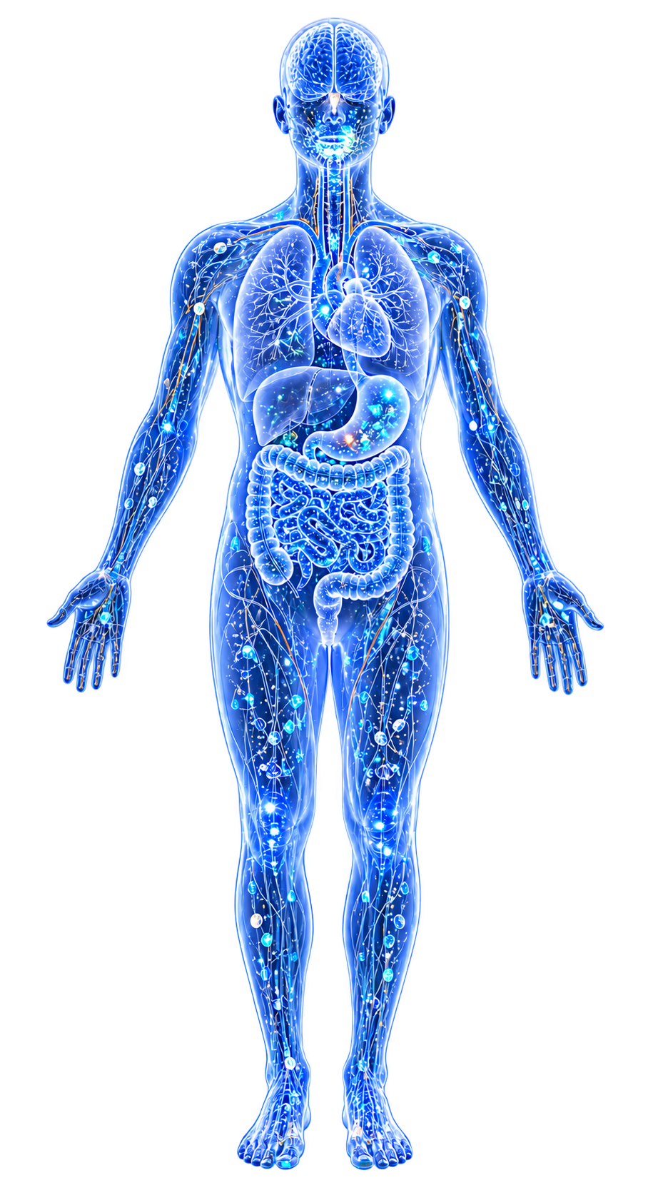
The pathological disruption of normal metal homeostasis—the tightly regulated balance of essential trace elements (Iron, Zinc, Copper, Manganese, Selenium, Cobalt) and the effective exclusion of toxic metals (Cadmium, Lead, Mercury, Arsenic).
Dyshomeostasis is a convergent mechanism underlying nearly every disease in this wiki and differs fundamentally from simple deficiency or toxicity: it is a failure of regulation, often occurring in the context of globally adequate metal levels but local or temporal mismanagement of distribution, speciation, or compartmentalization.
The distinction matters clinically. A patient can have normal serum iron and still have pathological iron accumulation in the substantia nigra. A patient can have normal plasma zinc and still have zinc-depleted immune cells.
Standard blood panels miss dyshomeostasis because they measure bulk systemic concentrations rather than the localized, compartment-specific metal distributions that drive disease.
Evidence map7 cited passagesInspect provenance +
Iron: Substantia nigra iron accumulation in parkinsons disease; cortical and hippocampal iron deposits in alzheimers disease; systemic iron overload in hereditary hemochromatosis. Excess iron drives ferroptosis—iron-catalyzed, lipid peroxidation-dependent programmed cell death.
Manganese: Occupational manganese exposure (welders, miners) causes manganism—parkinsonism from manganese accumulation in the globus pallidus and striatum.
Iron in anemia of chronic disease: Serum iron is low, but body iron stores (ferritin) are normal or elevated. The apparent "deficiency" is nutritional immunity—hepcidin-driven withholding of iron from circulation to deny it to pathogens. Treating this with iron supplementation worsens infection rather than correcting anemia.
A striking genetic demonstration of dyshomeostasis as a causal IBD mechanism comes from the Crohn's disease-linked ZIP8 A391T (rs13107325 in SLC39A8) variant:
The gut microbiome is both a target and a mediator of metal dyshomeostasis:
The bidirectionality is demonstrated by: toxic metals decrease microbial diversity and disrupt SCFA, bile acid, and amino acid metabolism, while microbiota actively methylate arsenic into less toxic forms, reduce mercury bioavailability, and produce metabolites that modulate metal absorption. The gut microbiome is not merely a passive victim of metal dyshome
| Source | Evidence Level | Key Contribution | |--------|---------------|-----------------| | (2024) | Animal model | Genetic proof of dyshomeostasis → dysbiosis → inflammation causal chain in IBD | | (2023) | Expert opinion (review) | Metal-specific dyshomeostasis patterns in AD and PD; therapeutic implications | | (2024) | Expert opinion (review) | Iron ho
Contents
1. What Dyshomeostasis Is Not2. Four Patterns of Dyshomeostasis3. The ZIP8-Microbiome Connection4. Metal-Specific Dyshomeostasis Profiles5. Dyshomeostasis as the Gut Microbiome Interface6. Key Studies7. Cross-ReferencesWhat Dyshomeostasis Is Not#
Not deficiency: Deficiency means inadequate total body content. Dyshomeostasis can coexist with normal or elevated total metal content but disrupted distribution.
Not toxicity (in the classical sense): Classical toxicology defines toxicity as excess above a threshold dose. Dyshomeostasis occurs at concentrations far below classical toxic thresholds—the harm comes from misregulation of normal levels, not from pharmacological overdose.
Not the same as Nutritional Immunity (Metal Sequestration): When the host sequesters metals in response to infection (hepcidin upregulation, calprotectin secretion, lactoferrin release), low serum metal levels reflect deliberate host defense—not dyshomeostasis. The key distinction is whether the metal redistribution is a regulated antimicrobial response or a pathological dysregulation.
Confusing the two leads to harmful interventions (e.g., iron supplementation during active infection).
Four Patterns of Dyshomeostasis#
1. Excess Accumulation#
Metal accumulates in tissue beyond functional capacity, generating reactive oxygen species via Fenton/Haber-Weiss chemistry and displacing other metals from their cofactor sites.
Iron: Substantia nigra iron accumulation in Parkinson's Disease; cortical and hippocampal iron deposits in Alzheimer's Disease; systemic iron overload in hereditary hemochromatosis. Excess iron drives Ferroptosis—iron-catalyzed, lipid peroxidation-dependent programmed cell death.[1]Common and Trace Metals in Alzheimer's and Parkinson's DiseasesDoroszkiewicz J, Farhan JA, Mroczko J et al. · 2023Open reference 1 ↓
Copper: Wilson's disease (ATP7B loss-of-function) causes hepatic and neurological copper accumulation that is directly neurotoxic; the disease model demonstrates that even an essential metal becomes pathological when redistribution fails.
Manganese: Occupational manganese exposure (welders, miners) causes manganism—parkinsonism from manganese accumulation in the globus pallidus and striatum.[1]Common and Trace Metals in Alzheimer's and Parkinson's DiseasesDoroszkiewicz J, Farhan JA, Mroczko J et al. · 2023Open reference 1 ↓
Cadmium: Cadmium accumulates in renal proximal tubular cells with a biological half-life of 10–30 years; it does not need to reach classically toxic concentrations to disrupt zinc-dependent enzymes in the kidney.
2. Local Depletion Despite Systemic Adequacy#
Metals are redistributed away from sites of functional need while overall body burden remains normal or elevated.
Zinc in immune cells: During acute phase response, zinc is redistributed from lymphocytes and macrophages to the liver for metallothionein synthesis. Immune cells become functionally zinc-depleted while serum zinc appears normal.
This impairs T-cell differentiation, reduces superoxide dismutase activity, and compromises antiviral defenses—without triggering any alarm on a standard blood panel.
Copper in cerebrospinal fluid: In both Alzheimer's and Parkinson's disease, CSF copper is reduced despite elevated brain copper at plaque and Lewy body surfaces—reflecting compartment-specific mislocalization rather than systemic copper deficiency.
Iron in anemia of chronic disease: Serum iron is low, but body iron stores (ferritin) are normal or elevated. The apparent "deficiency" is Nutritional Immunity (Metal Sequestration)—hepcidin-driven withholding of iron from circulation to deny it to pathogens.
Treating this with iron supplementation worsens infection rather than correcting anemia.[2]Mechanism of Iron Ion Homeostasis in Intestinal Immunity and Gut Microbiota RemodelingHonghong Bao, Yi Wang, Hanlin Xiong et al. · 2024Open reference 2 ↓
3. Ratio Disruption#
The relative balance between metals matters as much as absolute concentrations, because metals compete for the same transporters, binding sites, and regulatory circuits.
copper (Cu):zinc (Zn) ratio: Elevation of the copper-to-zinc ratio is documented across Colorectal Cancer, Cardiovascular Disease, polycystic-ovary-syndrome, and systemic Metal-Driven Inflammation. Elevated copper:zinc signals Oxidative Stress (copper-driven Fenton chemistry) combined with zinc depletion (reduced antioxidant defense via SOD1).
Toxic metal competition: Cadmium and lead compete with calcium, zinc, and iron for absorption via DMT1 and calcium channels (Mis-Metallation). When toxic metals are present, essential metals are displaced from their transporters, producing functional deficiency of essentials alongside toxic metal accumulation.
4. Temporal Dysregulation#
Homeostatic responses that are appropriate acutely become pathological when chronic.
Hepcidin-driven iron sequestration: Appropriate for days to weeks during acute infection. When sustained for months to years in chronic inflammatory disease (IBD, CKD, rheumatoid arthritis), it produces functional iron-restricted erythropoiesis and anemia of chronic disease—harming the patient while failing to resolve the underlying inflammatory stimulus.
Metallothionein upregulation: Protective acutely against cadmium, lead, or mercury exposure. Chronically maintained, it sequesters essential zinc in liver metallothionein at the expense of zinc availability to peripheral tissues.
The ZIP8-Microbiome Connection#
A striking genetic demonstration of dyshomeostasis as a causal IBD mechanism comes from the Crohn's disease-linked ZIP8 A391T (rs13107325 in SLC39A8) variant.[3]ZIP8 A391T Crohn's Disease-Linked Risk Variant Induces Colonic Metal Ion Dyshomeostasis, Microbiome Compositional Shifts, and InflammationYang JC, Zhao M, Chernikova D et al. · 2024Open reference 3 ↓
ZIP8 is a metal transporter expressed at the apical surface of intestinal epithelial cells. The A391T variant alters its transport kinetics.
Knock-in mice homozygous for the A391T variant show increased cobalt, cadmium, and manganese in mucosal tissue and reduced luminal availability of iron, cobalt, copper, zinc, cadmium, and manganese compared to wild-type.
This metal redistribution—more metal absorbed into the mucosa, less available in the lumen to gut bacteria—reshapes the Gut Microbiome composition in an age-dependent manner, with genotype accounting for 3% of microbiome variance at 2 months increasing to 9% at 12 months.
By 10 months, MUT mice develop spontaneous intestinal inflammation—absent at 5 months. The progression from metal dyshomeostasis to Dysbiosis to inflammation follows the temporal pattern expected if metal availability restructures the microbial community, which then drives inflammatory pathology.
This study establishes metal transporter polymorphism → metal dyshomeostasis → microbiome dysbiosis → inflammation as a causal chain in IBD—making dyshomeostasis not just a downstream consequence of disease but a primary upstream driver.
Metal-Specific Dyshomeostasis Profiles#
| Metal | Excess Pattern | Depletion Pattern | Key Disease Contexts |
|---|---|---|---|
| Iron | Ferroptosis, Fenton ROS, substantia nigra accumulation | Nutritional immunity (not true depletion) | Parkinson's, Alzheimer's, IBD |
| Copper | Wilson's disease, AD plaque copper, ceruloplasmin dysfunction | CSF copper depletion in neurodegeneration | Alzheimer's, Parkinson's, liver disease |
| Zinc | Rare—amyloid plaque zinc sequestration | Immune dysfunction, impaired SOD1, anorexia | Alzheimer's, IBD, chronic inflammation |
| Manganese | Manganism, globus pallidus accumulation | Rare in gut context | Parkinson's-like syndrome, occupational |
| Cadmium | Renal proximal tubule accumulation | Displaces zinc/calcium | CKD, IBD, cancer |
| Lead | Bone stores, BBB disruption | Displaces calcium, impairs development | Alzheimer's, developmental toxicity |
Dyshomeostasis as the Gut Microbiome Interface#
The gut microbiome is both a target and a mediator of metal dyshomeostasis.[4]Heavy Metal-Gut Microbiota Interactions: Probiotics Modulation and Biosensors DetectionLiliana Anchidin-Norocel, Oana C. Iatcu, Andrei Lobiuc et al. · 2025Open reference 4 ↓ Toxic metals (arsenic (As), cadmium (Cd), mercury (Hg), lead (Pb)) deplete SCFA-producing commensals while enriching metal-tolerant Proteobacteria, shifting the microbiome toward a dysbiotic state.
The dysbiotic microbiome then worsens metal handling: loss of commensal bacteria that normally biosorb, biotransform, or precipitate metals increases metal bioavailability; enrichment of siderophore-producing Proteobacteria increases iron competition; loss of Butyrate producers increases gut permeability, allowing greater metal absorption.
This creates a feedback loop: metal exposure → dysbiosis → impaired metal handling → more metal exposure → worse dysbiosis.
The bidirectionality is demonstrated by:[5]Toxic and Essential Metals: Metabolic Interactions with the Gut Microbiota and Health ImplicationsQinheng Zhu, Boyan Chen, Fu Zhang et al. · 2024Open reference 5 ↓ toxic metals decrease microbial diversity and disrupt SCFA, bile acid, and amino acid metabolism, while microbiota actively methylate arsenic into less toxic forms, reduce mercury bioavailability, and produce metabolites that modulate metal absorption.
The gut microbiome is not merely a passive victim of metal dyshomeostasis—it is an active participant in maintaining or failing to maintain metal homeostasis.
Key Studies#
| Source | Evidence Level | Key Contribution |
|---|---|---|
| [3]ZIP8 A391T Crohn's Disease-Linked Risk Variant Induces Colonic Metal Ion Dyshomeostasis, Microbiome Compositional Shifts, and InflammationYang JC, Zhao M, Chernikova D et al. · 2024Open reference 3 ↓ (2024) | Animal model | Genetic proof of dyshomeostasis → dysbiosis → inflammation causal chain in IBD |
| [1]Common and Trace Metals in Alzheimer's and Parkinson's DiseasesDoroszkiewicz J, Farhan JA, Mroczko J et al. · 2023Open reference 1 ↓ (2023) | Expert opinion (review) | Metal-specific dyshomeostasis patterns in AD and PD; therapeutic implications |
| [2]Mechanism of Iron Ion Homeostasis in Intestinal Immunity and Gut Microbiota RemodelingHonghong Bao, Yi Wang, Hanlin Xiong et al. · 2024Open reference 2 ↓ (2024) | Expert opinion (review) | Iron homeostasis regulation; nutritional immunity vs. true deficiency distinction |
| [5]Toxic and Essential Metals: Metabolic Interactions with the Gut Microbiota and Health ImplicationsQinheng Zhu, Boyan Chen, Fu Zhang et al. · 2024Open reference 5 ↓ (2024) | Expert opinion (review) | Bidirectional metal-microbiome interactions; ten metals mapped |
Cross-References#
- Mis-Metallation—toxic metals occupying essential metal cofactor sites
- Nutritional Immunity (Metal Sequestration)—regulated metal restriction as antimicrobial defense (not dyshomeostasis)
- Ferroptosis—iron-dependent cell death driven by iron dyshomeostasis
- oxidative stress—Fenton chemistry as primary ROS source from iron/copper excess
- Gut-Metal-Microbiome Interactions—the ecosystem where dyshomeostasis originates and is amplified
- Metal-Disease Matrix: A Cross-Source Synthesis—maps dyshomeostasis patterns across diseases
- inflammation—bidirectional relationship with metal imbalance
- Iron—the metal most extensively studied in homeostasis/dyshomeostasis
- Copper—Wilson's disease as the archetypal dyshomeostasis model
- Zinc—critical for immune function; redistribution during inflammation
- Parkinson's Disease—iron dyshomeostasis as a central driver
- Alzheimer's Disease—copper, zinc, and iron dyshomeostasis in plaque formation
References 6
Numbered by first appearance in the article, then reconciled with its declared source list.
- 1
Doroszkiewicz J, Farhan JA, Mroczko J et al. (2023). Common and Trace Metals in Alzheimer's and Parkinson's Diseases. International Journal of Molecular Sciences.
- 2
★Honghong Bao, Yi Wang, Hanlin Xiong et al. (2024). Mechanism of Iron Ion Homeostasis in Intestinal Immunity and Gut Microbiota Remodeling. International Journal of Molecular Sciences.
- 3
★Yang JC, Zhao M, Chernikova D et al. (2024). ZIP8 A391T Crohn's Disease-Linked Risk Variant Induces Colonic Metal Ion Dyshomeostasis, Microbiome Compositional Shifts, and Inflammation. Digestive Diseases and Sciences.
- 4
★Liliana Anchidin-Norocel, Oana C. Iatcu, Andrei Lobiuc et al. (2025). Heavy Metal-Gut Microbiota Interactions: Probiotics Modulation and Biosensors Detection. Biosensors.
- 5
★Qinheng Zhu, Boyan Chen, Fu Zhang et al. (2024). Toxic and Essential Metals: Metabolic Interactions with the Gut Microbiota and Health Implications. Frontiers in Nutrition.
- 6
★Giasuddin Ahmed, Md. Shiblur Rahaman, Enrique Perez et al. (2025). Associations of Environmental Exposure to Arsenic, Manganese, Lead, and Cadmium with Alzheimer's Disease: A Review of Recent Evidence from Mechanistic Studies. Journal of Xenobiotics.
Article network
Mentioned here 18
Pages linking here 6
Connect the evidence
Publicly readable discussion by ORCID-authenticated researchers. Questions, interpretation, methods, corrections, and new evidence stay attached to this record.
No discussion yet. Start with a precise question or a source-backed observation.
Activity and accepted changes
Accepted researcher context, editorial status, public discussion, and upstream Git revisions are shown together. Pending, declined, and withdrawn proposals remain private.
- published revision
Backfill oxidative stress concept links
Karen Pendergrass · +2 −2
Inspect exact Git diff ↗ - published revision
Backfill gut microbiome concept links
Karen Pendergrass · +1 −1
Inspect exact Git diff ↗ - published revision
Backfill inflammation concept links
Karen Pendergrass · +1 −1
Inspect exact Git diff ↗ - published revision
Complete corpus-wide Dysbiosis linking
Karen Pendergrass · +1 −1
Inspect exact Git diff ↗ - published revision
massive wiki expansion: 149 stubs fixed, 100+ new pages, Rule 15 scan, keystone papers
WikiBiome Deploy Bot · +20 −20
Inspect exact Git diff ↗ - published revision
pre-overnight checkpoint 2026-04-18
WikiBiome Deploy Bot · +1 −1
Inspect exact Git diff ↗ - published revision
Deepen 8 concept pages + 2 CP source pages (co-selection, AMR, alpha-synuclein, metabolites, gut-microbiome, propionic-acid, amyloid-beta, dyshomeostasis)
WikiBiome Deploy Bot · +99 −19
Inspect exact Git diff ↗ - published revision
Deep content + citation pass: 10 entities enriched, 7 new sources, DOI integrity
WikiBiome Deploy Bot · +1 −1
Inspect exact Git diff ↗ - published revision
WikiBiome update — integrity fixes, metallomic diet pages, cross-condition analyses
WikiBiome Deploy Bot · +3 −1
Inspect exact Git diff ↗ - published revision
WikiBiome update — 2026-04-10 23:44
WikiBiome Deploy Bot · +30 −0
Inspect exact Git diff ↗