
 metals · microbes · host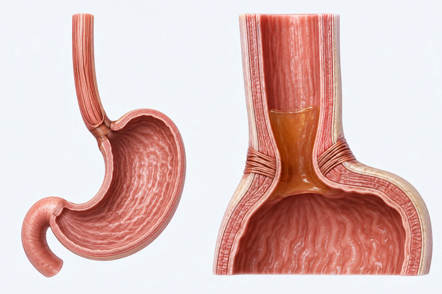
Representative gastroesophageal-junction anatomy with limited retrograde luminal flow. The fluid column is an editorial reconstruction, not a universal sphincter defect, mucosal injury, complication, severity measure, treatment response, or diagnosis.
Scientific media record1 verified identifier
- Subject
- Gastroesophageal Refluxcondition
- Identifiers
- MeSH:D005764
- Review
- Editorial review completeIdentifiers authority-verified · Accessibility validated · · gerd|gerd-pathology-v1.webp
- Digital source
- Trained-algorithmic mediaCreated with a trained generative algorithm and reviewed by WikiBiome for subject identity, scientific framing, identifiers, provenance, and accessibility.
- Scientific basis
- Gastroesophageal Reflux — MeSHDefinition & Facts for GER & GERD
- License
- CC BY-SA 4.0Created
Gastroesophageal reflux disease (GERD) is a chronic condition in which stomach acid or bile flows back into the esophagus, causing mucosal damage and symptoms such as heartburn and regurgitation. Affecting approximately 20% of Western populations, GERD is conventionally treated with proton pump inhibitors (PPIs), H2 blockers, and lifestyle modifications.
An emerging and underappreciated dimension of GERD involves nickel hypersensitivity, esophageal microbiome disruption, and the complex relationship between Helicobacter pylori colonization and reflux pathology. The PPI-microbiome axis further complicates the disease by introducing iatrogenic metal and microbial perturbations.
Evidence map32 cited passagesInspect provenance +
Mendelian randomization now provides causal-level evidence linking specific gut microbial taxa to GERD risk in both directions (n=78,707 GERD cases, 288,734 controls):
In reverse MR (GERD → microbiome), GERD itself had causal influence on 13 taxa:
Across 11 studies (2015–2025):
This dysbiotic shift activates TLR2/TLR4 signaling: TLR2 expression elevated 2.1-fold and claudin-1 (tight junction protein) decreased 47% in GERD patients with gram-negative dysbiosis, providing a direct molecular mechanism for barrier failure
SIBO prevalence significantly higher in GERD patients (P=0.007)
Oral microbiome in GERD patients is dominated by Prevotella and Haemophilus (n=266)—consistent with oral-esophageal translocation driving dysbiosis
Decreased Bifidobacterium: PPI-treated infants show Firmicutes increase to 65% relative abundance vs. Bifidobacterium dominance (72%) in controls; PPI duration correlated with alpha-diversity (r=0.42, P=0.01)
A critical and largely overlooked dimension of PPI therapy is its promotion of fungal dysbiosis (cross-sectional, n=65; gastric mucosal and fecal ITS sequencing):
| Observation | Evidence Summary | |-------------|-----------------| | Nickel sensitivity and GERD | A low-nickel diet improved symptoms in 95% of refractory GERD patients with confirmed nickel allergy | | PPI and microbiome disruption | Chronic PPI use is associated with Enterobacteriaceae expansion, CDI risk (1.5–2.7x), Bifidobacterium depletion, Candida c
Glutathione depleted: Glutathione metabolism pathway disrupted in GERD children (288 differential metabolites, ).
STEAP2 metalloreductase: Host SNPs in STEAP2 (iron/copper uptake enzyme) associated with esophageal microbiome composition.
The "cytokine sizzle" model:
| Marker | Direction | Evidence | |--------|-----------|---------| | TLR2 | 2.1-fold increase | Only gene significant after BH correction; LPS binds TLR2 on esophageal epithelium | | IL-6 | Elevated; decreased 38% by PPI | Driven by LPS-TLR2 activation; downregulates claudin-1 | | IL-8 | Elevated; decreased 41% by PPI | Correlated with esophageal Spirochaete
| Enriched in GERD/Barrett's | Evidence | Role | |---------------------------|---------|------| | prevotella (P. melaninogenica) | 4 studies; prevalence 22%→83% normal to metaplasia | Key Barrett's biomarker; distinct strain genomics in metaplasia | | Pseudomonas | Chen 2024 (62% vs 1.2%) | Massive enrichment in GERD/FED; LPS production | | veillonella | Des
Candida albicans detected in 96.9% of gastric mucosal samples. PPI treatment significantly increases Candida colonization. Fungal dysbiosis present in GERD regardless of PPI use; PPI further exacerbates it (, n=65).
The mechanistic pathway from dysbiosis to symptoms: Gram-negative bacteria produce LPS → LPS binds TLR2 (2.1-fold upregulation) → IL-6 secretion → claudin-1 downregulation (47%) → dilated intercellular spaces → submucosal sensory neuron exposure → symptoms. This pathway operates even when acid reflux is normal (functional esophageal disorder patients have th
Nickel (Ni)—The most striking metal connection. A low-nickel diet improved symptoms in 95% of refractory GERD patients regardless of patch-test nickel allergy status (19/20 patients; mean GERD-HRQL decrease of 27.05 points, P<0.001). Dietary nickel is abundant in legumes, nuts, whole grains, chocolate, and canned foods. Nickel-induced mast cell degranulati
Glutathione—Dysbiosis-associated altered glutathione metabolism documented in GERD.
Claudin-1—Tight junction protein decreased 47% in GERD patients with gram-negative dysbiosis (Chen et al. 2024); direct molecular evidence of barrier failure.
| Taxon | Role | Evidence | |-------|------|----------| | prevotella | Progressive enrichment: 22%→50%→58%→83% from normal to metaplasia; metaplasia strains carry TonBC iron transport and MlaD membrane domains |, | | veillonella | ↑52% in Barrett's/EAC; oral-esophageal translocation | | | fusobacterium nucleatum | Gram-negative anaerobe; LPS producer; oral o
| Taxon | Role | Evidence | |-------|------|----------| | streptococcus | Healthy esophageal dominant; ↓45% from BE to EAC |, | | lachnospiraceae UCG004 | Causally protective (MR OR=0.91); SCFA producer | | | actinobacteria | Phylum causally protective (MR OR=0.93) | | | akkermansia muciniphila | Strongest protective signal against Barrett's (MR OR=0.76) | |
Bidirectional MR (n=78,707 GERD cases, 288,734 controls) established that GERD and dysbiosis are causally linked in both directions:
Host genetics shape esophageal microbiome structure:
LPS biosynthesis—Gram-negative enrichment increases LPS load, activating TLR2/TLR4 signaling. TLR2 expression elevated 2.1-fold in GERD with gram-negative dysbiosis.
Showing 24 of 32 evidence-bearing passages. Every remaining citation is still indexed in the reference record below.
One disease. Five evidence layers.
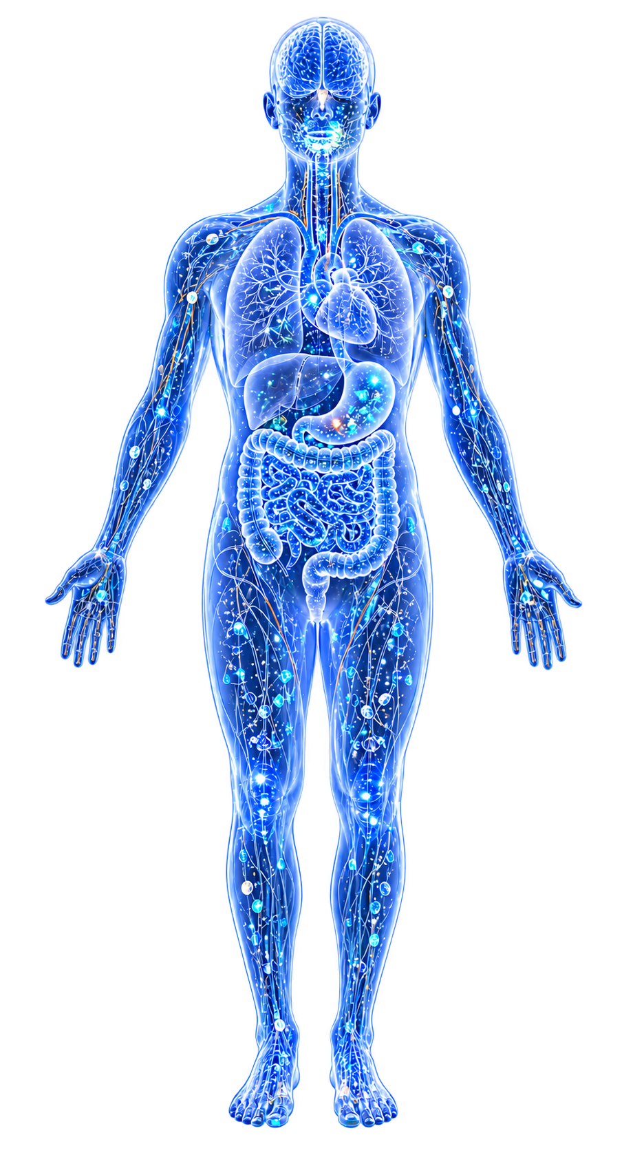
A generated systems view of the metals, organisms, host sequestration signals, ecological conditions, and microbial functions indexed for Gastroesophageal Reflux Disease (GERD).
Evidence layer
Taxonomic signature
Organisms reported as enriched or depleted, with their indexed functional context kept beside the name.Gram-negative anaerobe enriched in GERD esophagus; P. melaninogenica prevalence rises 22%→83% across reflux-metaplasia continuum; TonB_C iron transport domains in metaplasia strains
Dramatically enriched in GERD/FED esophagus (62% vs 1.2% in healthy); drives LPS-TLR2 activation
Gram-negative anaerobe enriched in GERD; ↑52% in Barrett's/EAC progression; oral-esophageal translocation
Key biomarker for Barrett's-to-EAC transition; ↑48% in progression; emerged in late-stage disease
F. nucleatum/necrophorum enriched; NF-kB correlated (gamma=0.68); cancer-associated
Expanded by PPI-driven pH elevation; LPS producers; causally increase Barrett's risk (OR=1.10 by MR)
Enriched in GERD gut; causal Barrett's risk factor by MR
B. stercoris/vulgatus/uniformis core enriched species; SIBO overlap
Fungal — detected in 96.9% of PPI-treated gastric mucosa; PPI-driven expansion reaches plateau within ~2 months; potential driver of persistent symptoms via visceral hypersensitivity
Gram-negative anaerobe; enriched in GERD and Barrett's; LPS-producing; oral origin
Causally increase GERD risk (OR=1.09) by Mendelian randomization
Enriched by GERD itself (reverse MR, OR=1.15); pro-inflammatory
Enriched by GERD (reverse MR, OR=1.24); pro-inflammatory
Dominant in healthy esophagus (Type I microbiome); ↓45% from BE to EAC; loss indicates ecological shift to gram-negative anaerobe dominance
Depleted by PPI therapy in infants (Firmicutes 65% vs. Bifidobacterium 72% dominance in controls)
Depleted in gut 16S data; paradoxically causal risk factor by MR — context-dependent
B. wexlerae/obeum depleted; SCFA producer loss
UCG004 causally protective (MR OR=0.91); SCFA producer; depletion removes barrier support
Causally protective against Barrett's esophagus (MR OR=0.76) — strongest protective signal identified
Phylum causally protective (MR OR=0.93); antibiotic production and immune modulation
Archaeon causally protective (MR OR=0.95); methane/SCFA ecology
Depleted by GERD itself (reverse MR, OR=0.85); health-associated lineage also depleted in obesity and metabolic syndrome
Evidence layer
Nutritional immunity
Host metal-withholding, inflammatory, antioxidant, and microbial-metabolite signals indexed in the signature.Elevated host signals
15Depleted protective signals
4Evidence layer
Ecological state
The environmental conditions that connect the organism-level observations into a system.Evidence layer
Virulence functions
Microbial structures, enzymes, and acquisition systems implicated by the linked evidence.The disease record, in full.
The original WikiBiome disease narrative remains intact beneath the generated signature atlas.
Metal Angle: Nickel Sensitivity#
The most striking metal connection in GERD is the role of dietary nickel in refractory disease. Yousaf et al. (2021) demonstrated that a low-nickel diet improved symptoms in 95% of refractory GERD patients who had confirmed nickel allergy via patch testing.
This finding reframes a substantial subset of treatment-resistant GERD as a manifestation of systemic Nickel Allergy and Allergic Contact Dermatitis rather than purely a motility or acid-secretion disorder.
Mechanisms of Nickel-Mediated GERD#
Dietary nickel is abundant in legumes, nuts, whole grains, chocolate, and canned foods—staples of many "healthy" diets. Systemic nickel allergy can trigger mucosal Metal-Driven Inflammation in the esophagus and stomach upon ingestion of high-nickel (Ni) foods. Nickel-induced mast cell degranulation in esophageal tissue releases histamine, which stimulates acid secretion.
The overlap between nickel sensitivity and eosinophilic esophagitis (EoE) may explain some refractory cases misdiagnosed as GERD. Patch testing for nickel allergy is not part of standard GERD workup, leading to chronic underdiagnosis.
Dietary Nickel Exposure#
Average dietary nickel intake ranges from 100-600 ug/day depending on food choices. Plant-based and whole-food diets tend to be higher in nickel, creating a paradox where "healthier" diets may worsen GERD in nickel-sensitive individuals. See Dietary Nickel Exposure for detailed food-level nickel content data.
Helicobacter pylori Relationship#
The relationship between H. pylori and GERD is controversial and paradoxical. H. pylori may be protective against GERD: Multiple epidemiological studies show an inverse relationship between H. pylori prevalence and GERD/esophageal adenocarcinoma rates.
H. pylori eradication has been associated with increased GERD symptoms in some patients, particularly those with corpus-predominant gastritis where the bacterium was suppressing acid secretion.
However, H. pylori is a clear risk factor for peptic ulcer disease and gastric cancer, creating a clinical dilemma. The "African enigma"—high H. pylori prevalence in Africa with low GERD rates—supports the protective hypothesis.
See Helicobacter pylori for the broader metal-microbiome interactions of this organism, including its nickel-dependent Urease and Hydrogenase enzymes.
Causal Microbiome-GERD Relationships#
Mendelian randomization now provides causal-level evidence linking specific gut microbial taxa to GERD risk in both directions[1]Wang K 2024 — Causal Gut Microbiota-GERD Associations via Bidirectional Mendelian RandomizationKui Wang, Suijian Wang, Yuhua Chen et al. · 2024Open reference 1 ↓ (n=78,707 GERD cases, 288,734 controls):
Protective Taxa (Causally Reduce GERD Risk)#
| Taxon | Level | OR (95% CI) | P-value | Mechanism |
|---|---|---|---|---|
| Clostridiales Vadin BB60 group | Family | 0.95 (0.91–0.99) | 0.027 | SCFA producer |
| Lachnospiraceae UCG004 | Genus | 0.91 (0.84–0.99) | 0.026 | Butyrate/SCFA producer |
| Methanobrevibacter | Genus | 0.95 (0.91–0.99) | 0.026 | Methane/SCFA ecology |
| Actinobacteria | Phylum | 0.93 (0.88–0.99) | 0.019 | Antibiotic production, immune modulation |
All four protective taxa are contributors to short-chain fatty acid (SCFA) biosynthesis. SCFAs (acetate, propionate, butyrate) are critical for colonocyte energy, intestinal barrier integrity, and motility regulation—directly relevant to lower esophageal sphincter (LES) function and mucosal protection.
Risk-Increasing Taxa (Causally Increase GERD Risk)#
| Taxon | Level | OR (95% CI) | P-value |
|---|---|---|---|
| Class Mollicutes | Class | 1.09 (1.01–1.19) | 0.037 |
| Genus Anaerostipes | Genus | 1.09 (1.01–1.16) | 0.017 |
| Phylum Tenericutes | Phylum | 1.11 (1.01–1.22) | 0.024 |
GERD Causes Dysbiosis: The Bidirectional Vicious Cycle#
In reverse MR (GERD → microbiome), GERD itself had causal influence on 13 taxa.[1]Wang K 2024 — Causal Gut Microbiota-GERD Associations via Bidirectional Mendelian RandomizationKui Wang, Suijian Wang, Yuhua Chen et al. · 2024Open reference 1 ↓
Depleted by GERD: Phylum Euryarchaeota (OR=0.82), Family Christensenellaceae (OR=0.85), Family Rikenellaceae (OR=0.88), Genus Rikenellaceae RC9 gut group (OR=0.78), Genus Ruminococcaceae NK4A214 (OR=0.89), Genus Anaerotruncus (OR=0.90), Genus Christensenellaceae R7 (OR=0.90).
Enriched by GERD: Collinsella (OR=1.15), Eggerthella (OR=1.24), Eubacterium rectale group (OR=1.12), Eubacterium ventriosum group (OR=1.12).
This bidirectional causality establishes GERD as both a consequence and a driver of Dysbiosis—a self-reinforcing loop where acid reflux depletes protective SCFA-producing communities, further impairing mucosal defense and perpetuating reflux.
Esophageal and Gut Microbiome Signature#
Across 11 studies (2015–2025):[2]Alageel 2025 — Examining the Microbiome Composition in Patients with Gastroesophageal Reflux Disease: A Systematic ReviewAlageel AA, Alomran DA, Alharbi HB et al. · 2025Open reference 2 ↓
Healthy vs GERD Microbiome#
Healthy esophagus: dominated by Streptococcus and gram-positive aerobes (Type I microbiome). GERD and Barrett's esophagus: shift toward gram-negative anaerobes—Veillonella, Prevotella, Fusobacterium, Neisseria (Type II microbiome).
This dysbiotic shift activates TLR2/TLR4 signaling: TLR2 expression elevated 2.1-fold and claudin-1 (tight junction protein) decreased 47% in GERD patients with gram-negative dysbiosis, providing a direct molecular mechanism for barrier failure.[2]Alageel 2025 — Examining the Microbiome Composition in Patients with Gastroesophageal Reflux Disease: A Systematic ReviewAlageel AA, Alomran DA, Alharbi HB et al. · 2025Open reference 2 ↓
Progression Signature: GERD → Barrett's → Esophageal Adenocarcinoma#
Lopetuso et al. (2020, n=26) documented the microbiome transition across disease stages. Streptococcus ↓45% from BE to EAC. Prevotella ↑60%, Veillonella ↑52%, Leptotrichia ↑48% in Barrett's and EAC.
Leptotrichia identified as key EAC biomarker—its enrichment may reflect the increasingly anaerobic, gram-negative ecology of pre-malignant esophageal tissue.
GERD and SIBO#
SIBO prevalence significantly higher in GERD patients (P=0.007).[2]Alageel 2025 — Examining the Microbiome Composition in Patients with Gastroesophageal Reflux Disease: A Systematic ReviewAlageel AA, Alomran DA, Alharbi HB et al. · 2025Open reference 2 ↓ Bacteroides uniformis (28%) and Bacteroides stercoris (22%) dominant in GERD+SIBO.
ABC transporter metabolism altered in SIBO-GERD overlap, with 288 differential metabolites including arachidonic acid pathway disruption—suggesting systemic metabolic consequences beyond the upper GI tract.
Oral-Esophageal Translocation#
Reflux events promote retrograde movement of gastric microbiota into the esophagus.
Oral microbiome in GERD patients is dominated by Prevotella and Haemophilus (n=266)—consistent with oral-esophageal translocation driving dysbiosis.[2]Alageel 2025 — Examining the Microbiome Composition in Patients with Gastroesophageal Reflux Disease: A Systematic ReviewAlageel AA, Alomran DA, Alharbi HB et al. · 2025Open reference 2 ↓
Periodontal pathogens detected in Barrett's esophagus tissue suggest chronic oral-esophageal microbial seeding.
PPI Effects on Gut and Fungal Microbiome#
PPIs are the most widely prescribed GERD therapy, but their effects extend far beyond acid suppression—including a profound and underappreciated fungal dimension.
Bacterial Effects of PPI#
Increased Enterobacteriaceae: PPIs reduce gastric acid barrier, allowing expansion of pathogenic gram-negative enteric bacteria. Increased Streptococcus: Oral streptococci colonize the gut when gastric pH is elevated.
Decreased Bifidobacterium: PPI-treated infants show Firmicutes increase to 65% relative abundance vs. Bifidobacterium dominance (72%) in controls; PPI duration correlated with alpha-diversity (r=0.42, P=0.01).[2]Alageel 2025 — Examining the Microbiome Composition in Patients with Gastroesophageal Reflux Disease: A Systematic ReviewAlageel AA, Alomran DA, Alharbi HB et al. · 2025Open reference 2 ↓
Increased CDI risk: PPI use associated with 1.5-2.7x increased risk of Clostridioides difficile infection. Metal absorption interference: PPIs reduce absorption of magnesium, calcium, iron, and zinc by raising gastric pH—potentially compounding metal deficiencies. SIBO risk: Small intestinal bacterial overgrowth more common in chronic PPI users.
Fungal (Mycobiome) Effects of PPI#
A critical and largely overlooked dimension of PPI therapy is its promotion of fungal dysbiosis[3]Shi 2023 — PPI-Induced Fungal Dysbiosis in Patients with Gastroesophageal Reflux DiseaseYichao Shi, Jianfeng Li, Shuntian Cai et al. · 2023Open reference 3 ↓ (cross-sectional, n=65; gastric mucosal and fecal ITS sequencing).
Candida detected in 96.9% of gastric mucosal samples from PPI-treated GERD patients (mean 43.59%, median 38.84%), significantly higher than untreated GERD and healthy controls.
PPI increases gastric mucosal Candida relative abundance significantly (P<0.05 vs. both nt-GERD and HC).
Plateau effect: No significant difference between short-term (average 6 months) and long-term (average 2 years) PPI users—Candida colonization reaches maximum within approximately 2 months.
Gastric mucosal fungal alpha diversity significantly reduced in PPI-treated patients (Shannon, Chao1; P<0.05 vs. HC).
Fecal mycobiome: Distinct from HC in GERD patients regardless of PPI status (ANOSIM R=0.12, P=0.004); Shannon diversity lower in untreated GERD vs. HC (P=0.021)—confirming a baseline fungal dysbiosis intrinsic to GERD itself.
Additional fungi enriched in PPI groups: Nothojafnea, Rhizodermea, Ambispora, Saccharicola (long-term PPI); Alternaria, Aspergillus, Mycenella, Exserohilum, Clitopilus (short-term PPI).
Mechanism: PPI-driven pH elevation removes the acid barrier that normally prevents Candida colonization, while simultaneously depleting acid-tolerant Lactobacillus species that competitively suppress Candida via displacement from epithelial surfaces.
Clinical implication: Candida dysbiosis may contribute to persistent symptoms in PPI-treated GERD through visceral hypersensitivity—consistent with evidence that intestinal fungal dysbiosis drives visceral hypersensitivity in IBS. This may explain why many long-term PPI users continue to report reflux symptoms despite adequate acid suppression.
Research Patterns and Observed Associations#
| Observation | Evidence Summary |
|---|---|
| Nickel sensitivity and GERD | A low-nickel diet improved symptoms in 95% of refractory GERD patients with confirmed nickel allergy[4]The effect of a low-nickel diet and nickel sensitization on gastroesophageal reflux disease: A pilot studyYousaf A, Hagen R, Mitchell M et al. · 2021Open reference 4 ↓ |
| PPI and microbiome disruption | Chronic PPI use is associated with Enterobacteriaceae expansion, CDI risk (1.5–2.7x), Bifidobacterium depletion, Candida colonization (96.9% in treated patients), and SIBO—effects extend to bacterial, fungal, and mineral absorption dimensions[3]Shi 2023 — PPI-Induced Fungal Dysbiosis in Patients with Gastroesophageal Reflux DiseaseYichao Shi, Jianfeng Li, Shuntian Cai et al. · 2023Open reference 3 ↓[2]Alageel 2025 — Examining the Microbiome Composition in Patients with Gastroesophageal Reflux Disease: A Systematic ReviewAlageel AA, Alomran DA, Alharbi HB et al. · 2025Open reference 2 ↓ |
| H. pylori and reflux | H. pylori eradication has been associated with increased GERD symptoms in some patients, particularly those with corpus-predominant gastritis where H. pylori was suppressing acid output |
| SCFA producers and GERD risk | Genetically higher abundance of Lachnospiraceae UCG004 and Clostridiales Vadin BB60 are associated with reduced GERD risk by MR[1]Wang K 2024 — Causal Gut Microbiota-GERD Associations via Bidirectional Mendelian RandomizationKui Wang, Suijian Wang, Yuhua Chen et al. · 2024Open reference 1 ↓ |
Associated Conditions#
GERD shares microbiome and ecological features with several related conditions:
| Condition | Shared Taxa | Shared Ecological Features |
|---|---|---|
| Barrett's esophagus | Prevotella ↑, Veillonella ↑, Leptotrichia ↑, Streptococcus ↓ | Gram-negative anaerobe dominance; LPS-driven inflammation |
| Esophageal adenocarcinoma | Leptotrichia ↑↑ (key EAC biomarker) | Progressive gram-negative enrichment; Streptococcus ↓45% |
| IBS | H. pylori overlap; altered Firmicutes/Bacteroidetes | Visceral hypersensitivity; SIBO; altered motility |
| SIBO | Bacteroides uniformis ↑, B. stercoris ↑ | Small intestinal bacterial overgrowth; ABC transporter metabolite disruption |
| Gastric cancer | H. pylori (driver); microbiome dysbiosis shared | Chronic mucosal inflammation; acid perturbation |
The GERD→Barrett's→EAC progression represents a well-documented example of how microbiome-driven inflammation can fuel oncogenic transformation—the Leptotrichia biomarker story being a potentially clinically actionable example.
Open Questions#
Unresolved questions identified by the current evidence record.
01What proportion of refractory GERD is attributable to undiagnosed nickel allergy?+
The current WikiBiome record identifies this as an unresolved evidence gap.
02Does nickel content in PPIs themselves (trace contamination) contribute to treatment failure in nickel-sensitive patients?+
The current WikiBiome record identifies this as an unresolved evidence gap.
03Can esophageal microbiome profiling predict progression from GERD to Barrett's esophagus—specifically, does Leptotrichia enrichment precede endoscopic changes?+
The current WikiBiome record identifies this as an unresolved evidence gap.
04Is H. pylori eradication net beneficial or harmful in GERD patients specifically?+
The current WikiBiome record identifies this as an unresolved evidence gap.
05Does treating SIBO in GERD patients improve reflux symptoms independent of acid suppression?+
The current WikiBiome record identifies this as an unresolved evidence gap.
06Does antifungal therapy in PPI-treated GERD patients improve persistent symptoms by reducing Candida-driven visceral hypersensitivity?+
The current WikiBiome record identifies this as an unresolved evidence gap.
07Can restoring SCFA-producing taxa (Lachnospiraceae UCG004, Clostridiales Vadin BB60) interrupt the GERD-dysbiosis self-reinforcing cycle identified by bidirectional MR?+
The current WikiBiome record identifies this as an unresolved evidence gap.
Connections#
- Nickel Allergy and Allergic Contact Dermatitis—Low-nickel (Ni) diet improved 95% of refractory GERD; systemic nickel allergy as overlooked GERD etiology
- Helicobacter pylori—Controversial protective relationship; nickel-dependent urease enzyme; protective against GERD through acid suppression in corpus-predominant gastritis
- Dietary Nickel Exposure—High-nickel foods as triggers; plant-based diet paradox in nickel-sensitive patients
- Probiotics—Potential to mitigate PPI-induced dysbiosis; restore SCFA-producing taxa
- dysbiosis—Esophageal microbiome shift from Type I to Type II; PPI-driven gut and fungal dysbiosis
- Actinobacteria (Actinomycetota)—Causally protective against GERD (OR=0.93); also protective against PPD and MDD—shared cross-condition pattern
- Lachnospiraceae—Causally protective (OR=0.91); SCFA production as mechanism; depleted across multiple GI diseases
- Candida albicans—PPI-promoted expansion in gastric mucosa (96.9% detection); potential driver of persistent symptoms through visceral hypersensitivity
- Barrett's Esophagus—Microbiome progression: Leptotrichia ↑48%, Streptococcus ↓45% from GERD to EAC
- sibo—Significantly associated with GERD (P=0.007); ABC transporter metabolite disruption in overlap
References 16
Numbered by first appearance in the article, then reconciled with its declared source list.
- 1
Kui Wang, Suijian Wang, Yuhua Chen et al. (2024). Wang K 2024 — Causal Gut Microbiota-GERD Associations via Bidirectional Mendelian Randomization. Frontiers in Immunology.
- 2
Alageel AA, Alomran DA, Alharbi HB et al. (2025). Alageel 2025 — Examining the Microbiome Composition in Patients with Gastroesophageal Reflux Disease: A Systematic Review. TPM (The Primary Care Companion for CNS Disorders).
- 3
Yichao Shi, Jianfeng Li, Shuntian Cai et al. (2023). Shi 2023 — PPI-Induced Fungal Dysbiosis in Patients with Gastroesophageal Reflux Disease. Frontiers in Cellular and Infection Microbiology.
- 4
Yousaf A, Hagen R, Mitchell M et al. (2021). The effect of a low-nickel diet and nickel sensitization on gastroesophageal reflux disease: A pilot study. Indian Journal of Gastroenterology.
- 5
Alnaim AA (2025). Effectiveness of dietary interventions in managing pediatric gastroesophageal reflux disease: a comprehensive systematic review. European Journal of Medical Research.
- 6
Liang T, Liu F, Liu L et al. (2021). Effects of Helicobacter pylori Infection on the Oral Microbiota of Reflux Esophagitis Patients. Frontiers in Cellular and Infection Microbiology.
- 7
Kucharczyk P, Parzecka KA, Symulewicz MJ et al. (2024). Innovative therapeutic strategies in the treatment of gastroesophageal reflux disease (GERD): A review of progress and perspectives. Wiadomosci Lekarskie Medical Advances.
- 8
Ye X, Yu F, Zhou J et al. (2023). Ye 2023 — Gut Microbiota in Children with GERD via Metagenomics and Metabolomics. Frontiers in Cellular and Infection Microbiology.
- 9
Deshpande NP, Riordan SM, Castano-Rodriguez N et al. (2018). Deshpande 2018 — Esophageal Microbiome Signatures and Host Genetics. Microbiome.
- 10
Park (2020). Park 2020 — NERD Treatment and Esophageal Microbiome. Scientific Reports.
- 11
Chen (2024). Chen 2024 — Esophageal Microbial Dysbiosis and TLR2 Signaling in GERD. Journal of Translational Medicine.
- 12
Luu (2022). Luu 2022 — Upper GI Microbiota in Children from Reflux to Metaplasia. Microbial Genomics.
- 13
Liu Y (2024). Liu Y 2024 — Bidirectional MR of Gut Microbiota with GERD and Barrett's Esophagus. BMC Genomics.
- 14
Gail A, Fero J, McCoy C et al. (2015). Bacterial Composition of the Human Upper Gastrointestinal Tract Microbiome Is Dynamic and Associated with Genomic Instability in a Barrett's Esophagus Cohort. PLoS ONE.
- 15
Sugihartono (2022). Sugihartono 2022 — Gastric Microbiota and H. pylori in GERD. Gut Pathogens.
- 16
Yin (2025). Yin 2025 — Probiotics Combined with PPI for GERD: Randomized Controlled Trial. Pharmacy Practice.
Article network
Mentioned here 14
Pages linking here 23
Connect the evidence
Publicly readable discussion by ORCID-authenticated researchers. Questions, interpretation, methods, corrections, and new evidence stay attached to this record.
No discussion yet. Start with a precise question or a source-backed observation.
Activity and accepted changes
Accepted researcher context, editorial status, public discussion, and upstream Git revisions are shown together. Pending, declined, and withdrawn proposals remain private.
- published revision
Backfill inflammation concept links
Karen Pendergrass · +1 −1
Inspect exact Git diff ↗ - published revision
Complete corpus-wide Dysbiosis linking
Karen Pendergrass · +3 −3
Inspect exact Git diff ↗ - published revision
Complete Hydrogenase contextual coverage
Karen Pendergrass · +1 −1
Inspect exact Git diff ↗ - published revision
Complete Tight junctions contextual coverage
Karen Pendergrass · +1 −1
Inspect exact Git diff ↗ - published revision
Complete reviewed Urease contextual coverage
Karen Pendergrass · +1 −1
Inspect exact Git diff ↗ - published revision
massive wiki expansion: 149 stubs fixed, 100+ new pages, Rule 15 scan, keystone papers
WikiBiome Deploy Bot · +34 −34
Inspect exact Git diff ↗ - published revision
cycle 1: health check + lint fixes + 8 ingests + 2 stubs + gestational-diabetes signature
WikiBiome Deploy Bot · +1 −0
Inspect exact Git diff ↗ - published revision
pre-overnight checkpoint 2026-04-18
WikiBiome Deploy Bot · +1 −1
Inspect exact Git diff ↗ - published revision
Batch: fix 1025 broken wikilinks, wire 13 STOP pages, deepen PPD/GERD/T1D/8 microbes
WikiBiome Deploy Bot · +9 −9
Inspect exact Git diff ↗ - published revision
Batch: fix 1025 broken wikilinks, wire 13 STOP pages, deepen PPD/GERD/T1D/8 microbes
WikiBiome Deploy Bot · +111 −31
Inspect exact Git diff ↗ - published revision
Deepen metal/concept entities + 8 new sources for T1D/schizophrenia
WikiBiome Deploy Bot · +233 −0
Inspect exact Git diff ↗ - published revision
v2 migration Priority 2: All 29 disease entity pages upgraded with associated_conditions, seo_target, wikipedia_differentiation
WikiBiome Deploy Bot · +6 −0
Inspect exact Git diff ↗ - published revision
WikiBiome v2 migration: signature pages + safety fixes + gap analysis
WikiBiome Deploy Bot · +9 −1
Inspect exact Git diff ↗ - published revision
WikiBiome update — 2026-04-11 22:49
WikiBiome Deploy Bot · +93 −0
Inspect exact Git diff ↗